Download
1 / 1
10 likes | 135 Views
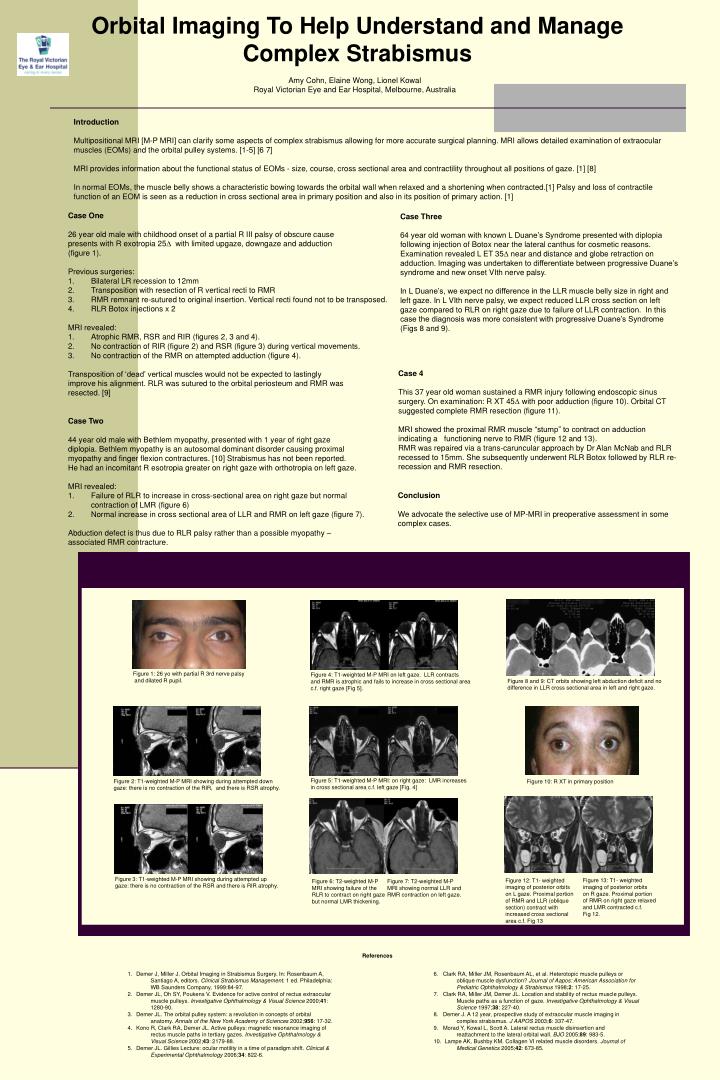
Figure 1: 26 yo with partial R 3rd nerve palsy and dilated R pupil. Case One 26 year old male with childhood onset of a partial R III palsy of obscure cause presents with R exotropia 25 with limited upgaze, downgaze and adduction (figure 1). Previous surgeries:

E N D
Figure 1: 26 yo with partial R 3rd nerve palsy and dilated R pupil. • Case One • 26 year old male with childhood onset of a partial R III palsy of obscure cause • presents with R exotropia 25 with limited upgaze, downgaze and adduction • (figure 1). • Previous surgeries: • Bilateral LR recession to 12mm • Transposition with resection of R vertical recti to RMR • RMR remnant re-sutured to original insertion. Vertical recti found not to be transposed. • RLR Botox injections x 2 • MRI revealed: • Atrophic RMR, RSR and RIR (figures 2, 3 and 4). • No contraction of RIR (figure 2) and RSR (figure 3) during vertical movements. • No contraction of the RMR on attempted adduction (figure 4). • Transposition of ‘dead’ vertical muscles would not be expected to lastingly • improve his alignment. RLR was sutured to the orbital periosteum and RMR was • resected. [9] Case Three 64 year old woman with known L Duane’s Syndrome presented with diplopia following injection of Botox near the lateral canthus for cosmetic reasons. Examination revealed L ET 35 near and distance and globe retraction on adduction. Imaging was undertaken to differentiate between progressive Duane’s syndrome and new onset VIth nerve palsy. In L Duane’s, we expect no difference in the LLR muscle belly size in right and left gaze. In L VIth nerve palsy, we expect reduced LLR cross section on left gaze compared to RLR on right gaze due to failure of LLR contraction. In this case the diagnosis was more consistent with progressive Duane’s Syndrome (Figs 8 and 9). Figure 2: T1-weighted M-P MRI showing during attempted down gaze: there is no contraction of the RIR, and there is RSR atrophy. Figure 3: T1-weighted M-P MRI showing during attempted up gaze: there is no contraction of the RSR and there is RIR atrophy. Figure 4: T1-weighted M-P MRI on left gaze. LLR contracts and RMR is atrophic and fails to increase in cross sectional area c.f. right gaze [Fig 5]. Figure 10: R XT in primary position Figure 5: T1-weighted M-P MRI: on right gaze: LMR increases in cross sectional area c.f. left gaze [Fig. 4] Figure 8 and 9: CT orbits showing left abduction deficit and no difference in LLR cross sectional area in left and right gaze. Figure 6: T2-weighted M-P MRI showing failure of the RLR to contract on right gaze but normal LMR thickening. Figure 7: T2-weighted M-P MRI showing normal LLR and RMR contraction on left gaze. Figure 13: T1- weighted imaging of posterior orbits on R gaze. Proximal portion of RMR on right gaze relaxed and LMR contracted c.f. Fig 12. Figure 12: T1- weighted imaging of posterior orbits on L gaze. Proximal portion of RMR and LLR (oblique section) contract with increased cross sectional area c.f. Fig 13 Orbital Imaging To Help Understand and Manage Complex Strabismus Amy Cohn, Elaine Wong, Lionel Kowal Royal Victorian Eye and Ear Hospital, Melbourne, Australia Introduction Multipositional MRI [M-P MRI] can clarify some aspects of complex strabismus allowing for more accurate surgical planning. MRI allows detailed examination of extraocular muscles (EOMs) and the orbital pulley systems. [1-5] [6 7] MRI provides information about the functional status of EOMs - size, course, cross sectional area and contractility throughout all positions of gaze. [1] [8] In normal EOMs, the muscle belly shows a characteristic bowing towards the orbital wall when relaxed and a shortening when contracted.[1] Palsy and loss of contractile function of an EOM is seen as a reduction in cross sectional area in primary position and also in its position of primary action. [1] Case 4 This 37 year old woman sustained a RMR injury following endoscopic sinus surgery. On examination: R XT 45 with poor adduction (figure 10). Orbital CT suggested complete RMR resection (figure 11). MRI showed the proximal RMR muscle “stump” to contract on adduction indicating a functioning nerve to RMR (figure 12 and 13). RMR was repaired via a trans-caruncular approach by Dr Alan McNab and RLR recessed to 15mm. She subsequently underwent RLR Botox followed by RLR re- recession and RMR resection. • Case Two • 44 year old male with Bethlem myopathy, presented with 1 year of right gaze • diplopia. Bethlem myopathy is an autosomal dominant disorder causing proximal • myopathy and finger flexion contractures. [10] Strabismus has not been reported. • He had an incomitant R esotropia greater on right gaze with orthotropia on left gaze. • MRI revealed: • Failure of RLR to increase in cross-sectional area on right gaze but normal • contraction of LMR (figure 6) • 2. Normal increase in cross sectional area of LLR and RMR on left gaze (figure 7). • Abduction defect is thus due to RLR palsy rather than a possible myopathy – • associated RMR contracture. Conclusion We advocate the selective use of MP-MRI in preoperative assessment in some complex cases. References • Demer J, Miller J. Orbital Imaging in Strabismus Surgery. In: Rosenbaum A, • Santiago A, editors. Clinical Strabismus Management. 1 ed. Philadelphia: • WB Saunders Company, 1999:84-97. • 2. Demer JL, Oh SY, Poukens V. Evidence for active control of rectus extraocular • muscle pulleys. Investigative Ophthalmology & Visual Science 2000;41: • 1280-90. • 3. Demer JL. The orbital pulley system: a revolution in concepts of orbital • anatomy. Annals of the New York Academy of Sciences 2002;956: 17-32. • 4. Kono R, Clark RA, Demer JL. Active pulleys: magnetic resonance imaging of • rectus muscle paths in tertiary gazes. Investigative Ophthalmology & • Visual Science 2002;43: 2179-88. • 5. Demer JL. Gillies Lecture: ocular motility in a time of paradigm shift. Clinical & • Experimental Ophthalmology 2006;34: 822-6. 6. Clark RA, Miller JM, Rosenbaum AL, et al. Heterotopic muscle pulleys or oblique muscle dysfunction? Journal of Aapos: American Association for Pediatric Ophthalmology & Strabismus 1998;2: 17-25. 7. Clark RA, Miller JM, Demer JL. Location and stability of rectus muscle pulleys. Muscle paths as a function of gaze. Investigative Ophthalmology & Visual Science 1997;38: 227-40. 8. Demer J. A 12 year, prospective study of extraocular muscle imaging in complex strabismus. J AAPOS 2003;6: 337-47. 9. Morad Y, Kowal L, Scott A. Lateral rectus muscle disinsertion and reattachment to the lateral orbital wall. BJO 2005;89: 983-5. 10. Lampe AK, Bushby KM. Collagen VI related muscle disorders. Journal of Medical Genetics 2005;42: 673-85.