Download
1 / 15
150 likes | 296 Views
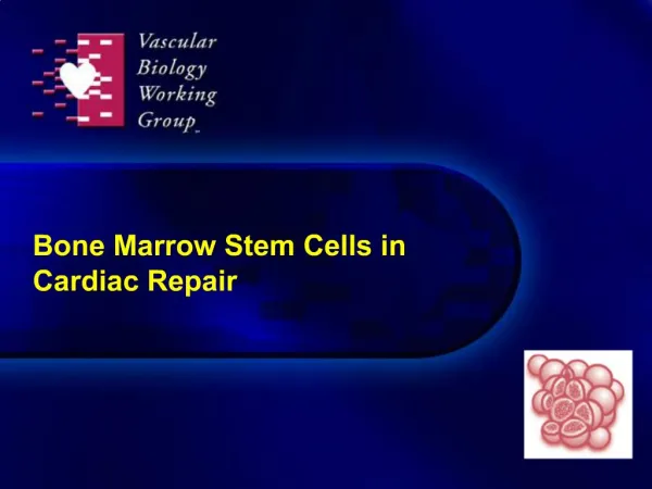
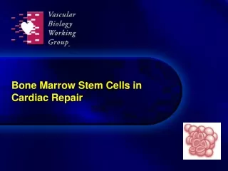
Bone Marrow Stem Cells in Cardiac Repair. Stem cells in cardiac repair: Proposed mechanisms of action. Cell homing and tissue integration. EC differentiation SMC differentiation. Cardiac differentiation fusion. Paracrine Effects. Attraction/ Activation of CSC. Angiogenesis.

E N D
Stem cells in cardiac repair: Proposed mechanisms of action Cell homing and tissue integration EC differentiation SMC differentiation Cardiac differentiation fusion Paracrine Effects Attraction/ Activation of CSC Angiogenesis Vasculo-genesis Arteriogenesis Cardiomyocyte proliferation Cardio-myogenesis ¯Cardiomyocyte apoptosis Modulation of inflammation Scar remodeling FUNCTIONAL IMPROVEMENT Dimmeler S et al. Arterioscler Thromb Vasc Biol. 2007;Oct. 19 epub.
Bone marrow cells promote myocardial regeneration: Postulated mechanism Infarcted myocardium Transplanted Cells Unknown molecular signal(s) Cell migration to damaged area Proliferation and differentiation Cytoplasmic proteins Nuclear proteins Cardian myosin α-Sarcomeric actin Connexin 43 Csx/Nkx2.5 MEF2 GATA-4 Functional competence Orlic D et al. Nature. 2001;410:701-5.
Cardiac stem cells are derived, in part, from bone marrow Post-mortem analysis of 4 hearts of female recipients of male BM transplants Demonstration of Y-chromosomes in up to 23% of cardiomyocytes Blue, green arrow = Y chromosome–positive true nucleus of BM Red = Derived cardiomyocyte cytoplasm (sarcomeric actin) surrounded by basement membrane laminin (green, arrowhead) Deb A et al. Circulation. 2003;107:1247-9.
Communication between heart and bone marrow signals in repair Heart endosteum Blood vessel endothelium SDF-1 SDF-1 transport CXCR4 Cell Recruitment Stem/progenitor cell Maturing leukocyte Blood vessel endothelium Bone marrow Bone Courtesy of Carl J. Pepine, MD
BMCs regenerate infarcted myocardium in mice Ventricular function LVDP LVEDP 120 100 80 60 40 20 0 40 30 20 10 0 * * † * † mm Hg mm Hg * LV +dP/dt LV –dP/dt 12000 8000 4000 0 12000 8000 4000 0 * † * † mm Hg s-1 mm Hg s-1 * * SO MI MI + BM SO MI MI + BM Orlic D et al. Nature. 2001;410:701-5.
BMCs reduce perfusion defect in ischemic pig hearts Kamihata H et al. Circulation. 2001;104:1046-52.
BMCs enhance collaterals to infarct region LAD Ligation BM-MNC after 3 weeks Kamihata H et al. Circulation. 2001;104:1046-52.
BMC therapy increases angiogenesis in ischemic pig hearts BM-MNC (Factor-VII) Control Medium (Factor-VIII) In part via enhanced synthesis of angiogenic factors in the infarcted region (ie, VEGF) Kamihata H et al. Circulation. 2001;104:1046-52.
Infarcted myocardium repair via autologous intracoronary mononuclear BMC transplantation Human model Strauer BE, et al. Circulation. 2002;106:1913-8.
BMCs minimize infarcted myocardium region 25 20 15 10 5 0 * P = 0.04 At 3 months: • SV index 49 56, P = 0.01 • Left ventricular end-systolic volume 8267, P = 0.01 • Thallium scintigraphy, cm2 174128 Infarct region (%) Cell therapy Standard therapy Strauer BE et al. Circulation. 2002;106:1913-8.
Assessment of intracoronary cell therapy in AMI PMC = peripheral mononuclear cells; RCT= randomized controlled trial; WMSI = wall motion score index. Lipinsky MJ et al. J Am Coll Cardiol.2007;50:1761-7.
Effects of intracoronary cell therapy on LVEF Study EF change % (random) EF change %or sub-category 95% CI 95% CI ASTAMI, 2005 -1.49 (-2.81, 0.01) Bartunek, 2005 -1.10 (-9.14, 2.94) BOOST, 2004 -2.83 (-3.00, -0.60) Jannsens, 2004 -1.10 (-2.68, 0.68) MAGIC-3, 2006 -2.20 (-7.18, 1.23) Meluzin, 2006 -2.03 (-2.94, -1.04) REPAIR-AM, 2006 -2.59 (-1.54, -1.44) Strauer, 2002 -1.03 (-4.06, 2.04) TCT-STAMI, 2006 -6.70 (-1.89, -3.51) Zhan-Quan, 2006 -5.50 (-3.19, -2.83) Total-2.97 (-1.04, -1.22) -10 -5 0 5 10 Favors cell therapy Favors control Test for heterogenicity, Chi2 = 33.62, af = 9 (P = 0.0001), P = 73.2%Test for overall effect: Z = 5.35 (P = 0.00001) Lipinsky MJ et al. J Am Coll Cardiol. 2007;50:1761-7.
Autologous CD34+ cells for intractable angina • N = 24 patients with CCS class 3/4 angina • G-CSF 5 μg/kg/day x 5 days • Leukapheresis performed on Day 5 • CD34+ cell selection • NOGA-guided transplantation to zones of myocardial ischemia • Phase I/IIa double-blind, 3:1 randomization, with crossover of placebo patients using frozen cells Losordo DW et al. Circulation.2007;115:3165-72.
Decrease in angina frequency with CD34+ cell therapy 3 6 Months Losordo DW et al. Circulation.2007;115:3165-72.