Download
1 / 11
110 likes | 324 Views
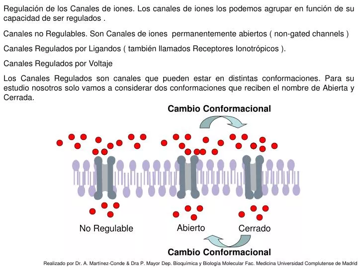
Regulación de los Canales de iones. Los canales de iones los podemos agrupar en función de su capacidad de ser regulados. Canales no Regulables. Son Canales de iones permanentemente abiertos ( non-gated channels ). Canales Regulados por Ligandos ( también llamados Receptores Ionotrópicos ).

E N D
Regulación de los Canales de iones. Los canales de iones los podemos agrupar en función de su capacidad de ser regulados . Canales no Regulables. Son Canales de iones permanentemente abiertos ( non-gated channels ) Canales Regulados por Ligandos ( también llamados Receptores Ionotrópicos ). Canales Regulados por Voltaje Los Canales Regulados son canales que pueden estar en distintas conformaciones. Para su estudio nosotros solo vamos a considerar dos conformaciones que reciben el nombre de Abierta y Cerrada. Cambio Conformacional Abierto No Regulable Cerrado Cambio Conformacional Realizado por Dr. A. Martínez-Conde & Dra P. Mayor Dep. Bioquímica y Biología Molecular Fac. Medicina Universidad Complutense de Madrid
Realizado por Dr. A. Martínez-Conde & Dra P. Mayor Dep. Bioquímica y Biología Molecular Fac. Medicina Universidad Complutense de Madrid
Realizado por Dr. A. Martínez-Conde & Dra P. Mayor Dep. Bioquímica y Biología Molecular Fac. Medicina Universidad Complutense de Madrid
Canales Regulados por ligandos. Son Canales que presentan diferentes conformaciones en presencia o ausencia de Ligando. Canal de iones que está Abierto en Reposo. Se Cierra al desaparecer el Ligando. Un ejemplo sería el Canal de Na+ y Ca++ de membrana de Bastones Retinianos, sensible a cGMP Cambio Conformacional El Canal en la célula en Reposo se encuentra en su conformación Abierta, La alta concentración citosólica de cGMP hace que este Ligando esté unido al Canal de Na+ y Ca++ y como consecuencia se encuentre estabilizado en su forma Abierta. La degradación del cGMP citosólico por el enzima fosfodiesterasa, causa un descenso en su concentración y como consecuencia una disociación del Canal. La disociación causa un cambio conformacional a la forma Cerrada. Al cesar el estímulo, y volver la célula a su estado de reposo, la concentración de cGMP se recupera, uniendose de nuevo al sitio alostérico del Canal. Pasando a la conformación Abierta, en la que queda estabilizado hasta que se produzca un nuevo estímulo. Cerrado Abierto Cambio Conformacional Realizado por Dr. A. Martínez-Conde & Dra P. Mayor Dep. Bioquímica y Biología Molecular Fac. Medicina Universidad Complutense de Madrid
La estructura de un canal de K+ • Bolas rojas grandes : agua / Bolas rojas pequeñas grupos C=0 cadena se señalan los aminácidos / Bolas verdes K+ • Close-up: Only three of the subunits are shown to provide a clearer view of the pore. All seven of the possible K+ locations are shown. However, only four, at most, are occupied at the same time; the other sites contain waters. Residues 75-79 comprise the selectivity pore and are shown as Sticks, colored CPK. The mainchain C=O's of these residues coordinate the K+ ions. The water-filled cavity is colored cyan. All of the waters shown here are highly-ordered and thus differ from the "bulk" water surrounding the channel. • K+ at sites 2 & 4: Each K+ ion is coordinated by eight oxygens with water between them. For example, K+ at site 4 is coordinated by the side chains and C=O's of Thr 75. • K+ between sites: The K+ ions are coordinated by only four of the mainchain C=O's. For example, the K+ between sites 2 and 3 is coordinated only by Val 76. A K+ is about to enter the pore from the cavity. • K+ at sites 1 & 3: The K+ ions are coordinated by eight mainchain C=O's. For example, K+ at site 1 is coordinated by Gly77 and Tyr78. The pore is normally occupied by two K+ ions and two waters. Concerted movement of the two ions and their waters leads to K+ exit from, or entry into the cell. The scripted animation shows the sequence of steps in the exit pathway. Realizado por Dr. A. Martínez-Conde & Dra P. Mayor Dep. Bioquímica y Biología Molecular Fac. Medicina Universidad Complutense de Madrid
Estructura del mismo canal de potasio en su forma abierta mostrando el paso del ión • Texto K+ Selectivity Pore • Close-up: Only three of the subunits are shown to provide a clearer view of the pore. All seven of the possible K+ locations are shown. However, only four, at most, are occupied at the same time; the other sites contain waters. Residues 75-79 comprise the selectivity pore and are shown as Sticks, colored CPK. The mainchain C=O's of these residues coordinate the K+ ions. The water-filled cavity is colored cyan. All of the waters shown here are highly-ordered and thus differ from the "bulk" water surrounding the channel. • K+ at sites 2 & 4: Each K+ ion is coordinated by eight oxygens with water between them. For example, K+ at site 4 is coordinated by the side chains and C=O's of Thr 75. • K+ between sites: The K+ ions are coordinated by only four of the mainchain C=O's. For example, the K+ between sites 2 and 3 is coordinated only by Val 76. A K+ is about to enter the pore from the cavity. • K+ at sites 1 & 3: The K+ ions are coordinated by eight mainchain C=O's. For example, K+ at site 1 is coordinated by Gly 77 and Tyr 78.The pore is normally occupied by two K+ ions and two waters. Concerted movement of the two ions and their waters leads to K+ exit from, or entry into the cell. The scripted animation shows the sequence of steps in the exit pathway. • K+EffluxMechanismThe steps shown in the animation (See Morais-Cabral et al., Figs. 4 and 5.): • State I Water-filled pore: The K+ that will soon exit is in the cavity. • State H K+ at site 4: The K+ enters the pore and is replaced by another in the cavity. • State E K+ at site 3: The K+ moves up one position (with the waters). Another K+ occupies the cavity from the cytosol. • State C K+ at sites 2 & 4: The K+ moves up another position. • Intermediate between site 2 and the extracellular site: The K+ is coordinated by only four of the mainchain C=O's prior to dissociation, first by Tyr 78 and finally by Gly 79. • Dissociation from extracellular site: The K+ binds briefly at site 0 and at the fully hydrated extracellular site, then diffuses into the extracellular solution. • Script ends with K+ at sites 2 & 4. For multiple cycles of ion efflux, the pathway resumes at State C above. K+ entry follows the same pathway in reverse direction. A single cycle takes about 10 nanoseconds to complete! Realizado por Dr. A. Martínez-Conde & Dra P. Mayor Dep. Bioquímica y Biología Molecular Fac. Medicina Universidad Complutense de Madrid
Realizado por Dr. A. Martínez-Conde & Dra P. Mayor Dep. Bioquímica y Biología Molecular Fac. Medicina Universidad Complutense de Madrid
Realizado por Dr. A. Martínez-Conde & Dra P. Mayor Dep. Bioquímica y Biología Molecular Fac. Medicina Universidad Complutense de Madrid
Canales Regulados por Ligandos Canal de iones que se Abre por interacción con un Ligando. Un ejemplo es el Canal de Na+ que es el Receptor Nicotínico de Acetil-Colina. Se encuentra en la membrana post-sináptica de nervio o placa motora. Cambio Conformacional El Canal en Reposo se encuentra en su conformación Cerrada, Cuando se degrada la Acetil-Colina de la hendidura sináptica, se producirá un descenso en la concentración de este neurotransmisor. Esto hace que se disocie del sitio de unión del Canal de Na+ y como consecuencia se induzca un cambio conformacional en el Canal que le hace pasar a la forma Cerrada de Reposo Un aumento en la concentración de Acetil-Colina en la hendidura sináptica hace que este neurotransmisor se una al sitio de unión del Canal de Na+ y como consecuencia se induzca un cambio conformacional en el Canal que le hace pasar a la forma Abierta Abierto Cerrado Cambio Conformacional Realizado por Dr. A. Martínez-Conde & Dra P. Mayor Dep. Bioquímica y Biología Molecular Fac. Medicina Universidad Complutense de Madrid
El Receptor Nicotínico de la Acetil-Colina Structure of the Nicotinic Acetylcholine Receptor PoreThe initial view of the AChR pore is from the synaptic (or extracellular space). Each subunit of the pentamer (a2bgd) is colored individually. The two a subunits are red. The four transmembrane helices are labeled at their N-termini in the g subunit (cyan). The central ion pore is formed by side chains from one side of helix M2.The membrane thickness indicated is the apolar region of the membrane. Side View: The protein is shown as Sticks from the plane of the membrane. The M2 helices are blue; the rest are red. In the intact AChR, the N-termini of each subunit connect to the ligand-binding domain (at the top). The width and height of the pore domain can be displayed (Å). • Pore Features • Pore Surface: The protein is shown Spacefill. One subunit has been removed to allow a view into the pore. The residues colored yellow form a "hydrophobic girdle" that is likely to have the gating function in AChR. The dark blue ball represents a Na+ ion trapped at the level of the maximal constriction. (The ion was inserted into the 1OED.pdb coordinate file for illustration.) • Pore Gate: The view is from the top of the pore. The M2 helices are shown as Backbone; each is labeled at the N-termini by subunit name. The hydrophobic girdle side chains are shown as Sticks, colored yellow; they are labeled on the a subunit. Note that although these residues are not identical in each subunit, they are similar in polarity and size. The "hypothetical Na+ ion" is shown as blue Spacefill. The gate in this channel is hypothesized to restrict passage of hydrated Na+ ions. Compare the structure shown here with that of say, the KcsA channel where main chain C=O groups interact with dehydrated K+ ions. The energetic cost of removing water with apolar side chains in this pore would be prohibitive. Realizado por Dr. A. Martínez-Conde & Dra P. Mayor Dep. Bioquímica y Biología Molecular Fac. Medicina Universidad Complutense de Madrid
Canal de iones que se Abre por interacción con una Proteína Reguladora. Un ejemplo sería el Canal de K+ de músculo cardiaco Regulado por las Subunidades Gibg de la Proteína Gi. Está regulado por el Receptor Muscarínico de de Acetil-Colina ACETIL-COLINA Transición Conformacional RECEPTOR GPCR MUSCARÍNICO K+ Realizado por Dr. A. Martínez-Conde & Dra P. Mayor Dep. Bioquímica y Biología Molecular Fac. Medicina Universidad Complutense de Madrid