Download
1 / 49
490 likes | 494 Views
This text explores how T cells recognize antigens, the concept of immunologic "self," and the role of major histocompatibility complex (MHC) molecules in graft compatibility and rejection. It also discusses the mechanisms of T cell clonal recognition and the impact of MHC polymorphisms on the immune response.

E N D
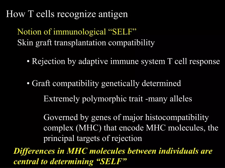
How T cells recognize antigen Notion of immunological “SELF” Skin graft transplantation compatibility • Rejection by adaptive immune system T cell response • Graft compatibility genetically determined Extremely polymorphic trait -many alleles Governed by genes of major histocompatibility complex (MHC) that encode MHC molecules, the principal targets of rejection Differences in MHC molecules between individuals are central to determining “SELF”
Clonal recognition of antigen by T lymphocytes • Each clonal T cell receptor (TCR) is specific for a particular sequence of amino acids in a peptide • The peptide is generated from proteins in the peptide-presenting cell • The peptide is not directly identified by the TCR, but recognized in a form bound to a MHC molecule
Clonal recognition of antigen by T lymphocytes The huge diversity of pathogen peptides to be recognized mandates a large number of different T cell clones, necessitating: • A somatic recombination mechanism to generate the large number of clones • A clonal selection process to identify the appropriate clones for the clonal repertoire
(Self-peptides are used as a surrogate for foreign peptides since there are few non-self peptides) *** The selection process (Thymic “education”) has two stages • First stage selects clones capable of recognizing self peptide in an individual’s own MHC molecules - positive selection • Second stage eliminates overtly self reactive clones with high affinity for self peptide:MHC- negative selection
Polymorphic residues of MHC The TCR repertoire differs from individual to individual • The specificity of self/non-self peptide binding to MHC molecules determined by pockets that only bind certain amino acid side chains • MHC genes are extremely polymorphic and alleles encode pockets with specificities for different amino acid side chains The TCR is specific for both peptide and MHC- A complex ligand The definition of immunologic self is made by selecting the clonal T cell repertoire on self-peptides bound to the individual’s particular allelic forms of MHC molecules
Immunologic self is both the collective recognition specificity of an individual’s T cell repertoire for peptides presented by the MHC and the set of self-peptides and self-MHC molecules that generated the repertoire
The immune response to pathogens Many of the events in the clonal selection process are recapitulated during the clone’s response to peptide in an immune response Non-self peptides from pathogens, etc., are bound to MHC molecules and recognized by T cells because their peptides are partial mimics of self Polymorphic residues of MHC
Because of MHC polymorphisms we each likely recognize non-self antigens and pathogens differently from the next individual Major selective advantages to the species However because the adaptive immune system is patterned on self, it sets the stage for the development of autoimmune disease
T cells recognize peptide antigens from two main types of pathogens Bacteria or components of an extracellular pathogen that have been internalized Virus - or Pathogen - infected cell B cell Any nucleated cell Macrophage/DC A viral peptide on a cell signifies it is infected and should be killed, while a pathogen’s peptide on a phagocytic cell signifies the cell has ingested a foreign substance and must be helped to eliminate the pathogen The adaptive immune system must make this distinction
The immune system makes this distinction by loading and recognizing peptides in either class I or class II MHC Cytosolic Virus or Pathogen Ingested Bacteria or Endocytic Pathogen Extracellular Pathogen or Toxin Challenge: Presenting cell: Any cell Macrophage/DC B cell Cytosol MHC class I CD8 T cells Death of cell presenting the viral antigen Endocytic vesicles MHC class II(or I) CD4 T cells (or CD8) Activation of cell to enhance pathogen killing Peptide degraded in: Peptides bind to: Presented to: Effect on presenting cell of T cell recognition: Endocytic vesicles MHC class II CD4 T cells Provision of help to B cell for production of antibodies
Structure of MHC molecules What are the structural features of the MHC that determine peptide binding?
Organization of the MHC Two classes of MHC molecules 0 1 2 3 4m bp Class II loci Class I loci HLA-DR HLA-DQ HLA-DP HLA-A HLA-B HLA-C Human Leukocyte Antigens (HLA) Ch 6
a1 a2 N Structure of peptide-binding class I MHC domain
The ligand for the CD8 T cell TCR Peptide-binding domains b2m MHC Class I Domains
The overall structure of class I and class II MHC is rather similar Class I Class II
MHC class I and II molecules have homologous domain organization, but different chain structure Peptide a1 a2 a1 b1 b2m a3 a2 b2 MHC class I molecule MHC class II molecule
The Structure of MHC Molecules: MHC Class I • The a chain is~ 350 AA long • Three globular domains, a1, a2 and a3, each ~90AA • a1 and a2 form the antigen-binding cleft • b2 microglobulin ~100AA, associates with the a3 domain, not MHC encoded • Transmembrane and cytoplasmic portion
The Structure of MHC Molecules: MHC Class II • Composed of two similar membrane spanning proteins, the a-chain and b-chain both encoded within the MHC • Each chain is made of two globular domains, each ~90AA, e.g. a1 and a2 • a1 and b1 domains form the antigen-binding cleft
At the genetic level the MHC class I and II system is complex! Two levels of diversity: Within individual: duplicated loci Class I: HLA-A, HLA-B, HLA-C Class II: HLA-DR, HLA-DQ, HLA-DP Between individuals: hundreds of alleles for most of these loci The MHC is by far the most genetically polymorphic region in the genome
A B A (isoforms or isomorphs) A1 A1 B1 A2 A2 B2 Alleles (Allotypes) In different individuals A3 A3 B3 A4 B4 A4 The diversity of MHC class I and II genes An evolutionary response to the structural diversity and mutation potential of microorganisms Arises from two mechanisms: Duplication of a gene locus in an individual resulting in multiple loci, polygeny Development of multiple alleles at a locus among individuals in the species, polyallelism
MHC alleles have a dominant mode of action because the molecules they encode have the ability to bind particular peptides Corollary: if your MHC molecules cannot bind a peptide, you cannot make a T cell response against it Duplicating a locus allows it to mutate and develop new peptide presenting specificities that operate in parallel with older ones
However each duplication increases “self” and mandates more negative clonal selection during repertoire formation, reducing the size of the repertoire Practical maximum is ~ three loci each for class I and class II HLA-DR HLA-DQ HLA-DP HLA-A HLA-B HLA-C No limit on the number of alleles
How T cells recognize antigen Notion of immunological “SELF” Skin graft transplantation compatibility • Rejection by adaptive immune system T cell response • Graft compatibility genetically determined Extremely polymorphic trait -many alleles Governed by genes of major histocompatibility complex (MHC) that encode MHC molecules, the principal targets of rejection Differences in MHC molecules between individuals are central to determining “SELF”
Clonal recognition of antigen by T lymphocytes • Each clonal T cell receptor (TCR) is specific for a particular sequence of amino acids in a peptide • The peptide is generated from proteins in the peptide-presenting cell • The peptide is not directly identified by the TCR, but recognized in a form bound to a MHC molecule
Clonal recognition of antigen by T lymphocytes The huge diversity of pathogen peptides to be recognized mandates a large number of different T cell clones, necessitating: • A somatic recombination mechanism to generate the large number of clones • A clonal selection process to identify the appropriate clones for the clonal repertoire
(Self-peptides are used as a surrogate for foreign peptides since there are few non-self peptides) *** The selection process (Thymic “education”) has two stages • First stage selects clones capable of recognizing self peptide in an individual’s own MHC molecules - positive selection • Second stage eliminates overtly self reactive clones with high affinity for self peptide:MHC- negative selection
Polymorphic residues of MHC The TCR repertoire differs from individual to individual • The specificity of self/non-self peptide binding to MHC molecules determined by pockets that only bind certain amino acid side chains • MHC genes are extremely polymorphic and alleles encode pockets with specificities for different amino acid side chains The TCR is specific for both peptide and MHC- A complex ligand The definition of immunologic self is made by selecting the clonal T cell repertoire on self-peptides bound to the individual’s particular allelic forms of MHC molecules
How peptides bind to class I MHC molecules How does polymorphism influence binding of different peptides and what is its immunologic significance?
How peptides bind Cluster of tyrosines recognize NH2 Class I MHC COOH NH2 + charged amino acids interact with negative COOH Specific bound peptide always oriented in the same direction in the MHC Length of peptide is constrained to 8 or 9 amino acids
How peptides bind 3 5 7 N C peptide 9 2 MHC class I TCR Anchor Anchor Rules for binding to MHC class I molecules • Usually peptides are 8-9 amino acids in length • Always oriented with NH2 terminus to the left • Most often anchored by interactions of the side chains of their 2nd (P2) and 9th (P9) amino acids to MHC pockets that confers specificity for amino acids with similar physical properties, e.g. size, charge, hydrophobicity, etc.
Y L Y L Y I Pr 1 Different Proteins yield different peptides that can bind to the same MHC molecule because they share anchor residues Y=Tyr L=Leu I=Ile Pr 2 Pr 3 Each different MHC Class I allotype has pockets that preferentially bind different amino acid side chains at positions P2 and P9 Only a few peptides from each protein will bind to a given MHC class I allotype
2 5 7 8 peptide 1 4 6 9 Protein 1 Protein 2 Protein 3 Peptide binding to class II MHC molecules Similar pockets for peptide side chains, but peptides binding class II molecules vary in length 12-24 AA and are anchored in the middle
For both class I and class II molecules • Binding is highly stable with no peptide interchange on cell surface • Specificity for original loaded peptide is maintained because empty MHC molecule is very unstable
How are proteins metabolized within the cell to yield peptides that bind to the MHC? How do cytosolic peptides from protein synthesized in virally infected cells get loaded on class I and not class II molecules to trigger killing by CD8T cells? How do peptides from ingested proteins get loaded on class II and not class I molecules to elicit macrophage activation and B cell help?
Class I peptide loading Synthetic Cytosolic peptides synthesized by virally infected cells get loaded on class I MHC endocytic The endocytic and synthetic pathways are usually quite separate
Peptides generated in the cytosol by the proteasome bind to MHC class I molecules while they are being synthesized within the ER
Top Side
The 26S proteasome is composed of the 20S proteasome core containing the catalytic chamber and two 19S regulatory complexes that bind and unfold ubiquitylated proteins 3 of the 7 subunits in each b ring of the 20s core confer the proteolytic activity, b1, b2 and b5 Responsible for the proteolysis of polyubiquitylated proteins in basal protein turnover
Proteasome core catalytic chamber cross section showing a ring of 7 b subunits Three have proteolytic activity; b1, b2 and b5 Proteins are unfolded and pass through the active proteolytic sites
IFN-g greatly enhances antigen processing and presentation • An immunomodulatory cytokine produced by activated T helper lymphocytes, CD8 CTLs and natural killer (NK) cells • Upregulates classic MHC I and II and TAP gene products • Upregulates the synthesis of three new proteasome subunits with different proteolytic activity that replace the three constitutively active proteolytic subunits, changing specificity of proteasome towards production of hydrophobic peptides that have greater binding affinity for MHC pockets
Generation of the immunoproteosome 3 immunosubunits LMP2 (ib1), LMP7 (ib5) and MECL-1 (ib2) replace the constitutive b1, b5 and b2 subunits in the 20S core and change specificity to more hydrophobic peptides
Generation of peptides for class I MHC requires precise proteolysis because the overall peptide length is restricted to 8 or 9 residues The 26S immunoproteasome system is used to get precise cuts at the C termini of the peptides Other peptidases nibble back the N termini until the peptide fits exactly, e.g. the IFN-g-inducible cytosolic leucine aminopeptidase (LAP) cleaves a single residue from the N- terminus The process has a relatively low efficiency, especially since the peptide production system is not coordinated with the peptide binding specificity of the individual’s MHC class I molecules
Almost no peptides presented by class I molecules are derived from “old” proteins A proportion of newly synthesised proteins are defective ribosomal products (DRiPs) that are rapidly ubiquitylated and degraded by proteasomes DRIPS are the major source for the generation of antigenic peptides
synthetic Class II peptide loading pathway Endocytic Proteins endocytized by DC, B cell or macrophage are contained in endocytic vesicles
Class II presentation is centered in the vesicular system Acidic endosomal proteases digest ingested proteins into peptides that will load MHC class II molecules This process does not require the precise proteolysis needed in the class I system, since the peptide terminii are not constrained by MHC class II
Invariant chain (Ii) A chaperone that complexes with MHC class II molecules during their synthesis in the endoplasmic reticulum has two critical actions • Ii Blocks the class II peptide binding groove and prevents loading by peptides destined for class I molecules • The recognition sequence on the Ii transmembrane portion targets the nascent MHC II molecule from the synthetic compartment to the acidic endosomal compartment to join with the degraded ingested exogenous peptides
Ii is degraded to CLIP (Class II-associated invariant chain peptide) by specific endosomal acidic cysteine proteases (cathepsins)
CLIP is only bound to the MHC groove by its peptide backbone and its side chains do not engage the pockets • HLA-DM, an ancient but non-classical class II molecule catalyzes the release of CLIP and the binding of high affinity peptides via interaction of peptide amino acid side-chains with MHC pockets • Without Ii the MHC class II molecule traffics to the cell membrane