Download

1 / 85
870 likes | 1.13k Views
The Genitourinary System. Chapter 12. All the best Santas are furry. The Genitourinary System. Includes animal reproduction and the processing of liquid waste…. The Urinary System. This system includes the organs that produce and excrete the waste substance urine. These organs are:

E N D
The Genitourinary System Chapter 12
The Genitourinary System Includes animal reproduction and the processing of liquid waste…
The Urinary System • This system includes the organs that produce and excrete the waste substance urine. • These organs are: • Kidneys • Ureters • Bladder • Urethra
Feline Urinary System Female Male
The Kidneys (nephro or reno) • Bean shaped organs located on both sides of the vertebral column • Retroperitoneal - Situated outside the peritoneal cavityRenal fasciae surround the kidneys, holding them in place. • Hilus – • A concave depression on each kidney’s medial margin • Provides an entrance for blood vessels, nerves and the ureter
The kidneys are dark reddish brown (except in felines, which are yellowish red). A cross section shows the external cortex and the internal medulla.
Kidney Transplantation Recipient Donor
The Urinary System • Renal sinus – Cavity containing the renal pelvis, blood vessels and fat • Renal pelvis - A reservoir that occupies most of the sinus and funnels urine to thr ureter • Ureter - The outlet tube that connects the renal pelvis & bladder
Medullary pyramids make up the medulla, & stud the walls of the renal sinus. Urine collects through ducts.
The nephron is the functional unit of the kidney, consisting of the renal corpuscle and renal tubule. There are about 1 million nephrons in the kidney. The Nephron
The renal corpuscle consists of a double walled cup shaped structure called the glomerular or Bowman’s capsule, which contains a twisted cluster of capillary channels called the glomerulus.
. The Kidney The kidney functions as a filter for waste products in the blood, excreting them in the urine. These waste products include nitrogenous wastes from the breakdown of proteins, toxins , mineral salts, excess glucose and water.
Urinary System: The Kidney(Video) http://www.youtube.com/watch?v=zEpUQkQ-uKM
Blood Pressure & The Kidneys • The speed that blood filters through the kidneys is affected by blood pressure (BP). • If systemic BP drops, as in shock, blood filtration may slow to a point where the kidneys stop functioning. • If BP is too high, kidney damage may result.
The kidneys affect the rate of secretion of some hormones, synthesize other hormones and maintain the pH of the blood so it remains neutral.
The Ureters • Muscular tubes extending from the kidneys to the bladder • Walls of the ureters are made up of: • An outer fibrous tissue layer • Two central layers of smooth muscle • Mucous membrane lining
The Ureters • Enter the neck of the bladder obliquely at the trigone • The flow of urine back to the kidney is effectively controlled by a natural valve
Urine • Enters the bladder every 10-30 seconds in spurts, rather than a continuous flow • Spurts are produced by peristaltic waves. • Ureteral opening on bladder opens every 2-3 seconds, then closes until another peristaltic wave opens it again • Prevents urine from flowing back into the ureters during bladder contraction
The Urinary Bladder • An elastic sac lying in the pelvis • Formed of 3 layers of smooth muscle lined with mucous membrane • Size & position depends on how much urine it contains • Has 2 openings to receive urine from the ureters, and another opening into the urethra
At the junction of the neck of the bladder with the urethra is a sphincter muscle that controls the amount of urine that passes into the urethra. • The bladder has two main functions: • Storing urine • Excreting urine
The voiding of urine from the bladder is called micturition. The act of preventing or concluding urination is a learned and voluntary action in more intelligent forms of animal life.
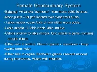
The Urethra • A membranous tubular canal that carries urine from the bladder to the exterior of the body • In the female, urethral length varies by species • The exterior opening of the urethra is called the urinary meatus
In the female the urinary meatus is located between the vagina and the clitoris directly cranial to the vulva. The only function of the urethra is urination.
In the male, the urethra varies in length by species and penile structure. • It is narrower than the female, so more prone to blockages from urolithiasis (stone formation). • It extends from the neck of the bladder through the accessory sex glands, between the fascia and through the penis.
The Male Urethra • Has 3 sections: • Prostatic • Membranous • Cavernous • The exterior opening is called the urinary meatus and serves a dual function - carrying both urine and reproductive secretions.
Normal Urine • A clear, pale amber color in most species, with a characteristic odor. • Is about 95% water • Contains dissolved substances such as nitrogenous waste, electrolytes, toxins, pigments, hormones, and abnormal substances like glucose, albumin or blood
Average urine output in a 24 hour period varies by: • Species • Temperature • Water intake • Type of work the animal is performing
Male Reproductive Organs • The basic male reproductive organs (gonads) are the testes • The ducts are the epididymides, vas deferentia, ejaculatory ducts and urethra
Accessory sex glands (not present in all species) include the seminal vesicles, prostate, bulbourethral and coagulating glands. The penis, scotum and spermatic cord are the primary reproductive structures.
The Testes • A pair of egg-shaped glands normally located in a sac-like structure called the scotum. • Size, shape, and location vary depending on species. • Each testicle is enclosed in a fibrous, white capsule called the tunica albuginea.
. The Testes • The testes have two functions: • Producing spermatozoa • Secreting hormones • Sperm cells are produced by the seminiferous tubules. The primary hormone, testosterone, is secreted by the cells of Leydig.
Testosterone has several functions: • Induces/maintains male secondary sex characteristics • Massive head/shoulders, crest of withers, tusks on boars, horns on rams • Influences muscle and bone growth
Male Female
Male animals generally have less subcutaneous fat, and the meat is less tender and juicy. Castration is performed on all animals intended for meat production to eliminate accumulation of testosterone after maturity.
Testosterone also influences: • Fluid & electrolyte metabolism • Has an excitatory effect on the kidney tubule • Suppresses anterior pituitary secretions
Epididymus • A pair of tightly coiled tube-like structures • Acts as a place for sperm to mature and stores sperm before ejaculation • Secretes a small portion of the seminal fluid