Download
1 / 24
260 likes | 566 Views
Chapters 12 Motor System – Cerebellum. Chris Rorden University of South Carolina Norman J. Arnold School of Public Health Department of Communication Sciences and Disorders University of South Carolina. Function of Cerebellum. Error Control Device - Monitor, Quality Control

E N D
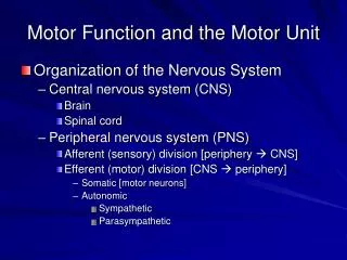
Chapters 12 Motor System – Cerebellum • Chris Rorden University of South Carolina Norman J. Arnold School of Public Health Department of Communication Sciences and Disorders University of South Carolina
Function of Cerebellum • Error Control Device - Monitor, Quality Control • Monitors outputs to muscles from motor cortex and sensory signals from receptors • Compares the efferent project plan with execution at motor action site • Considers related factors and makes adjustments
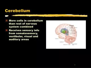
Cerebellum • 50% of brain’s neurons, 10% of volume • Can change movements as necessary • E.G. Walking or talking • Does not reach conscious awareness • Muscle synergy or coordination monitored • Important in running, speaking - all fluid movements
tentorium cerebelli • "tent of the cerebellum" • dura mater that separates the cerebellum from the inferior portion of the occipital lobes.
Posterior Cranial Fossa • Fossa is a depression or cavity in the bone • Cerebellum, pons, and medulla oblongata sit in the Posterior cranial fossa
Cerebellar Anatomy Seen from feet • Located dorsal to pons and medulla • In posterior fossa under tentorium cerebelli • Lobes • Floccular Nodular(small fluffy mass) • Anterior • Posterior Anterior lobe (H) Posterior lobe (I)
Flattened Cerebellum • Longitudinally separated into hemispheres and cortices • Median (Vermal) Vermis=worm • Paramedian (Paravermal) • Lateral
Cerebellum Paramedian Primary Fissure Median Posterior Superior Fissure Horizontal Fissure Prepyramidal Fissure Posterolateral Fissure
Cerebellar Nuclei (Nuclei = deep cluster of neurons) • Dentate nucleus • Largest, communicates through cerebellar peduncle • Carries information important for coordination of limb movements (along with the motor cortex and basal ganglia) • Emboliform nucleus (medial side of the nucleus dentatus) • Regulates movements of ipsilateral extremity • Globose nucleus • Regulates movements of ipsilateral extremity • Fastigial nucleus • Regulates body posture • Is related to the flocculo nodular lobe
Dentate Nucleus Dentate Nucleus Pontine Projections Superior Cerebellar Peduncle Pons
Somatotopic Organization • Tactile information • Ipsilateral anterior lobule • Bilateral paramedian lobules • Cerebral Cortex and Cerebellum have similar representations • Motor representation • Same area as sensory mapping • May have auditory and visual processing
Transverse Cerebellar Regions • Floccular nodular lobe (Archicerebellum ) • Oldest, related to vestibular part of CN VIII • Regulates equilibrium through vestibulospinal tract • Anterior lobe (Paleocerebellum) • Rostral to Primary Fissure • General Sensory Receptors • Concerned with muscle tone and walking • Posterior lobe (Neocerebellum) • Newest and Largest, Receives afferent projections from contralateral sensorimotor cortex • Projects to contralateral motor cortex • Functions in coordination of fine and skilled movements
Longitudinal Cerebellar Regions • Vermis • Contributes to body posture • Paravermal region • Regulates movements of ipsilateral extremities (e.g. walking) • Lateral Zone • Regulates skilled movements of ipsilateral extremity (e.g. tying your shoe)
Cerebellar Connection • Three Peduncles • Inferior – afferent: mediate sensorimotor input to the cerebellum • Middle – afferent: same as above • Superior – efferent: transmit output from the cerebellum to the brainstem and on to the thalamus, motor cortex, and spinal cord • Varied afferents to Cerebellum : • spinal cord • brainstem • motor cortex • Afferenet:Efferent Ratio = 40:1 • For each going from cerebellum to body, 40 coming in
Afferent Pathways (Inferior) • Vestibulocerebellar Tract • Info From Semicircular Canals Through Inferior Peduncle • Maintains Upright Posture • Dorsal Spinocerebellar Tract • Info From Reticular Nuclei (involved in regulation of sleep, respiration, heartbeat, etc.) • Unconscious Proprioception From Muscle Spindles, Golgi Tendons and Tactile Receptors
Afferent Pathways (Inferior 2) • Reticulocerebellar Tract • Info From Cerebral Cortices, Spinal Cord, Vestibular Complex, and Red Nucleus • Olivocerebellar Tract • Info From Spinal Cord Through Olivary N to Contralateral Cerebellar Hemisphere • Source of Climbing Fibers for Direct Input to Cerebellum • Cuneocerebellar Tract • Mediate Proprioception From Upper Limbs and Neck
Afferent and Efferent Projections Thalamus Red nucleus Superior Cerebellar Peduncle Middle Cerebellar Peduncle (pontocerebellar fibers) Inferior Cerebellar Nucleus (olivocerebellar fibers)
Afferent Pathways (Middle) • Info From Pontine Nuclei From Opposite Cerebral Cortex, Visual and Auditory Inputs • To Opposite Cerebellar Hemisphere
Efferent Pathways • Arise From Cerebellar Nuclei • Dentate nucleus • Emboliform nucleus • Globose Nucleus • Through Superior Cerebellar Peduncle to • Red Nucleus (Brainstem) • Thalamus • Motor Cortex
Cerebellar Cortex • Structured in Three Parallel Layers • Molecular • Purkinje • Connecting Surface and Deep Cerebellar Nuclei • Source of All Efferent Fibers • Cerebellar Cortex • Granular • Have Mossy Fiber Axons to Purkinje Axons
Clinical Considerations • Signs of Dysfunction • Impaired Muscle Synergy • Reduced Muscle Tone • Evident in Skilled Tasks • Ataxia • Lack of Order and Coordination in Activities • Slow Movement (Bradykinesia) • Mild Muscular Weakness (Asthenia) • Asynergia (Poor coordination of muscles: Dysdiadochokinesia) • Speech difficulties (Ataxic Dysarthria) • affects respiration, phonation, resonance and articulation, but most pronounced in articulation and prosody.
Clinical Considerations 2 • Dysdiadochokinesia • Clumsiness in Alternating Movements • Switching between supination and pronation – e.g. screwing in a light buld • Tapping, Speech Sound • Dysarthria • Ataxic Dysarthria • Poor articulation: Slurred and Disjointed Speech • Dysmetria • Error in Judgment of Range and Distance of Target • Undershooting or Overshooting
Clinical Considerations 3 • Intentional Tremor • Accessory Movement During Volitional Task • vs. Parkinson’s Disease Where Tremor Lessens During Volitional Movement • Hypotonia • Reduced Resistance to Passive Stretch • Rebounding • Inability to Predict Movement • Cannot Hold Back Movement • Disequilibrium • Unsteady Gait, Body Wavering
Cerebellar Pathologies • Cerebrovascular Accident (CVA) • Thrombotic, embolic or hemorrhagic • Vertebrobasilar Artery • Toxicity • Chronic Alcoholism • Progressive Cerebellar Degeneration • Friedrich's Ataxia: Autosomal Recessive Heredity Degenerative Condition • Combined Sensory and Motor Dysfunctions • Poor coordination of Gait and speech