Download
1 / 34
340 likes | 477 Views
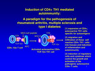
Induction of CD4+ TH1 mediated autoimmunity: A paradigm for the pathogenesis of rheumatoid arthritis, multiple sclerosis and type I diabetes. (1) expansion of CD4+, autoreactive TH1 cells specific for autoantigens

E N D
Induction of CD4+ TH1 mediated autoimmunity: A paradigm for the pathogenesis of rheumatoid arthritis, multiple sclerosis and type I diabetes (1) expansion of CD4+, autoreactive TH1 cells specific for autoantigens (2) migration and infiltration of these self reactive CD4+ TH1 cells into tissues and induction of inflammation and autoimmunity (3) induction of regulatory cells and cytokines which control the growth and activation of the pathogenic autoreactive CD4+ T cells MHC/self-peptide CD4 CD4 MHC/Vb TCR Vbx TCR Vbx APC CD4+ Vbx T cell Activated autoreactive CD4+ TCR Vbx TH1 cell
Rheumatoid Arthritis: Definition Rheumatoid arthritis is characterized by a chronic inflammation of the synovial joints and infiltration by blood-derived cells, chiefly memory T cells, macrophages, and plasma cells, all of which show signs of activation. This leads in most cases to progressive destruction of cartilage and bone, which occurs after invasion of these tissues by the cellular synovial tissue and is believed to be mainly mediated by cytokine induction of destructive enzymes, chiefly matrix metalloproteinases. There is also prominent development of new vessels and evidence of systemic inflammation, for example, upregulated acute phase proteins. In more severe cases there is involvement of vessels and other organs.
Rheumatoid Arthritis: Genetics Twin and other genetic studies have demonstrated that a major genetic contribution to disease predisposition resides in the MHC class II HLA-DR locus. Females are about 2-3 times more susceptible than males. More than 80% of caucasian rheumatoid patients express DR1 or DR4 subtypes which share an epitope mapping to amino acids 70-74 of the DRß chain, in the polymorphic region lining the peptide binding groove. There is recent evidence that the genetically susceptible HLA-DR4 (e.g., DR0401) alleles bind different peptides in their peptide binding groove than the non-susceptible (e.g., DR0402) alleles. Susceptible alleles bind a negatively charged amino acid at the p4 pocket of the binding groove. Mutation analysis revealed that position 71 of the DRß chain in particular correlates with the genetic linkage of RA susceptibility
Shared epitope in RA Amino acid sequences in the b chain HLA-DRB*0401 molecules dictate susceptibility to RA
Clinical Manifestations of Rheumatoid Arthritis (1) Arthritis (a) Symmetrical involvement of the small joints of the hands and feet, particularly the proximal interphalangeal (PIP), metatarsophalangeal (MTP), and metacarpophalangeal (MCP) joints, but involvement of wrists, ankles, knees, elbows, and hips is also common. (b) When the disease involves the axial skeleton, it is most frequently in the cervical region.
Clinical Manifestations of Rheumatoid Arthritis (3) Associated Syndromes (a) Sjogren’s syndrome-salivary gland inflammation and keratoconjunctivitis (b) Felty’s syndrome-profound neutropenia, thrombocytopenia and splenomegaly (C) Amyloidosis-type II
Villous Synovitis Rheumatoid Synovium (Pannus) Normal synovium Pannus Invading and Destroying cartilage and Bone
Initiating Event in Rheumatoid Arthritis CD2 DR4/peptide a,b TCR Fc Receptor a,b CD4 CD4 T cell Macrophage/ dendritic cell/B cell
The Initiating Event in Rheumatoid Arthritis RA auto antigen SmIg or FcR APC CD40 ER Golgi CD3 a,b TCR HLA-DM &DO DR4/ peptide endosome DR4/CLIP CD4 CD28 DR4/ InvChain CD4 T cell CD80 (B7.1) The putative RA inducing protein binds antigen specific SmIg on B or phagocytic receptors on dendritic cells cells and is internalized and digested into peptides in endosomes and bound to the MHC class II molecule (DR4). The DR4/peptide complex triggers the TCR on antigen specific T cells leading to T cell activation.
RA auto-antigenic peptide DR4, DR1
Th0 CD4+ T Cells Differentiate into Distinct Th1 and Th2 Subsets Cytokine Profile Functions T-macrophage interactions DTH responses Intracellular pathogens IL-2, IFN-g, TNFa GM-CSF, LT Th1 IL-12 IFN-g + - IL-10 IFN-g - T-B cell interactions Antibody responses Extracellular pathogens IL-4 + IL4, IL-5, IL-6 IL-10, IL-13 Th2
Consequences of CD40L/CD40 interactions on Dendritic cell function MHC class II/ autopeptide CD3 TCR CD40 B7 Activated T cell CD40 CD40L CD40 CD28 B7 MHC class II (1) induction of cytokines (IL-8, IL-12, TNF-a, MIP-1) (2) stimulation of B71 and B72 expression and co-stimulatory function with activation of T cell growth (3) augmentation of antigen-presenting function
T-Macrophage Interactions Induce Synovial Cell Proliferation and Activation DR4/peptide CD2 a,b, TCR Fc Receptor CD4+ TH1 Cell CD4 Rheumatoid factor (RF) CD40L Macrophage IL-1, TNF, TGFb BONE RESORBTION CD40 inflammation Synthesis of PGE-2, Collagenase, IL-1 Synovial Cell Proliferation
Final Phases of B cell Differentiation are Mediated by Contact T cell signals (CD40L/CD40) and Lymphokines CD23 CD40L TCR Sm Ig CD4 CD40 Activated T cell Activated B cell L ymphokines IL-4, IL-5, IL-6, IFN-g, TGFb IgG IgA IgE Plasma Cell
Inflammation Immunopathophysiology of Rheumatoid Arthritis Dendritic cell/ APC CD2 Activated TH1 CD4+ T Cell CD4+ Cell (TH0 ) IL-12 DR4/RA peptide CD40L a,b, TCR IFN-g CD40 IL-4 RF Macrophage Sm Ig CD4+ Cell (TH2 ) Fc R PGE-2, collagenase chemokines RA antigen IL-1, TNF, TGFb B Cell Osteoclast activation Synovial fibroblast Endothelial cell Rheumatoid factor (RF) Plasma Cell (1) Synthesis of PGE-2, Collagenase, IL-1 (2) synovial cell proliferation Bone and cartilage destruction hypothalamus Vasculitis Fever
Rheumatoid Factors • (1) Characteristics of RF's • (a) RF's are autoantibodies with specificity for the Fc region of self-IgG • (b) Most RF's are IgM but IgG and IgA RF's are also observed • (2) Biologic Occurrence and Disease Associations • (a) RF's are the major autoantibodies observed in RA • (b) RF's can be induced in experimental animals by injection of either denatured IgG or by immunization with bacteria. • (c) High titered RF is seen in chronic inflammatory conditions such as rheumatoid arthritis, other rheumatic conditions, TB and SBE • (3) Biologic and Pathologic Functions of RF's • (a) RF's may play a role in augmenting the phagocytosis of opsonized particles and in the clearance of immune complexes. • (b) RF bound to IgG or to immune complexes can precipitate in vessel walls and induce vasculitis. High titered RF is associated with systemic vasculitis in RA • (c) Rheumatoid factors can bind to Fc receptors on macrophages and augment the release of monokines, including IL-1 and and TNF
Rheumatoid Factors and Immune Complexes Augment the Activation of Macrophages DR4/peptide CD2 a,b, TCR Fc Receptor CD4+ TH1 Cell Rheumatoid factor (RF) or Immune complexes CD4 CD40L Macrophage IFN-g IL-1 CD40 Increased synthesis of IL-1, TNF, TGFb, IL-6, PGE-2 and Collagenase Rheumatoid factor (RF) Activated Macrophage
TNF, IL-1 and RANK-L activate osteoclasts to induce bone resorption CD2 a,b, TCR Activated CD4+ T Cell Mf/dendritic cell RANK-L CD40 TNF CD40 Soluble RANK-L B7 MHC class II TNFR Soluble RANK Receptor RANK TNF IL-1 PGE2 Precursor Osteoclast Bone Resorption Activated Osteoclast
Mechanisms of action of drugs used to treat RA (a) Block T-APC interaction antibodies to MHC class II, CD4 or the TCR (b) Decrease T cell activation cyclosporine, anti-CD3, anti-CD28, anti-CD80 (B7), anti-CD40L, CTLA-4 agonist (e) Inhibit products of T/macrophages NSAIDs, TNF receptor inhibitors, IL-1 receptor inhibitors (c) Prevent T cell, B cell or synovial cell proliferation Methotrexate, immuran, cytoxan (d) Inhibit T cell or APC function steroids, gold, penicillamine
CD3 z z g d e h h a b New Surface Molecules are Expressed after T cell Activation CD45RA CD2 CD4 TCR CD28 T Cell Activation CD3 z z g d e h h MHC Class II CD25 IL-2R VLA-1 a b F AS L CD45 RA CD45 RO CD40L CD2 CD28 CD4 TCR CTLA-4
TNF IL-1