Download
1 / 15
150 likes | 347 Views
Calcium Imaging of Dorsal Root Ganglion Cells using Fura-2 AM ( acetoxy-methyl-ester). What is Dorsal Root Ganglion (DRG)?.

E N D
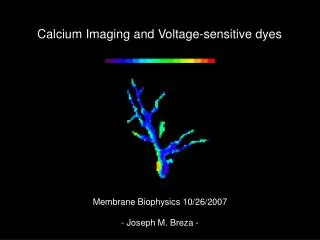
Calcium Imaging of Dorsal Root Ganglion Cells using Fura-2 AM (acetoxy-methyl-ester).
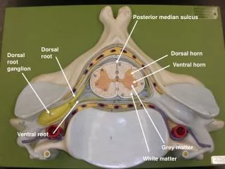
What is Dorsal Root Ganglion (DRG)? It is a collection of cell bodies of sensory neurons associated with the spinal nerves lying along the vertebral column of the spine. The cell body of each sensory neuron in the dorsal root ganglion has a long axon which extends from the dendrites located in the skin (receptors), muscles, tendons, joints and internal organs to the spinal cord then heads towards the brain. These dendrites monitor touch (various kinds), stretch, temperature, and pain. DRG cells here are used to screen for anti-pain activity of compounds isolated from bacteria of marine snails. spinal cord
Why calcium imaging? Ca2+ is a universal second messenger involved in specific cellular processes and regulation including muscle contraction, fertilization, synaptic transmission, cell division, blood clotting etc.). Changes in intracellular Ca2+ concentration are often within millisecond and can not be visualized directly in living cells. Thus, specific molecules (e.g. Fura-2) with optical properties that change upon interacting with Ca2+ are used to monitor changes in Ca2+ concentration inside the cells. Fura-2 is one of the most common calcium indicators is which it has an emission peak at 505 nm and changes its excitation peak from 340 nm to 380 nm in response to calcium binding, allowing the concentration of intracellular calcium to be determined from the ratio of fluorescence emission or excitation at distinct wavelengths. The main advantage of using Fura-2 dye from other probes is that the ratio signal is independent of the dye concentration, illumination intensity, and optical path length. Here presents the intracellular calcium elevations in neurons and other excitable cells in dorsal root ganglion cells using Fura-2.
euthanize and dissect 2 to 3-week old C57/B6 (male) mouse remove DRG into 1x HBSS solution, treat with trypsin for 20 min, then wash with MEM 3X triturate to dissociate cells, preplate for 1-3 hr collect cells and centrifuge at 8,000 rpm for 5 min seed neurons in poly-L-lysine and laminin coated 24-well plate incubate at 37oC, 5% CO2 for 18-24 hours load cells with Fura-2 fluorescent dye in the dark for 40-60 min wash cells 3X with oxygenated observation medium, pH 7.4 with final volume of 500 uL calcium image the cells treated with samples in an oxygen-supplied chamber analyze graph images Methodology 1 animal dissection 2 DRG cell culture 3 Calcium imaging
340 nm brightfield with marked cells 380 nm control panel brightfield events list cell response Calcium imaging window
control well / KCl control treated wells 2 increased second KCl response/ inhibitory 1 KCl/wash wash 2 KCl/wash 1 KCl/wash extract/drug KCl+drug/wash List of events for imaging
Activity profiles of interests 1 decreased second KCl response/ inhibitory 2 increased second KCl response/ excitatory 3 with drug response and increased second KCl response
Interesting Results CT8: low 2nd KCl response in 30% of the cells CT8: no drug response in 10% of the cells
CT4: low 2nd KCl response 1-18-4: slightly low 2nd KCl response in less than 10% of the cells
106T: slightly high 2nd KCl response 1-18-7: low 2nd KCl response
1-18-6 (1): with drug response; most of the cells not able to recover after KCl+drug treatment 1-18-6 (1): with drug response and higher 2nd KCl response
CN48: high 2nd KCl response 1-18-6 (2): higher 2nd KCl response
CN127: with drug response but similar 1st and 2nd KCl responses
Summary of results • Thirty samples were tested, some were done in duplicates. Seven of them showed activity at low ug/mL on DRG cells.
Additional Info • Will use DRG cell line to speed up screening. F11 cell line will be sent this week (c/o Alan Light). • Response of 3-5% population of neurons should be included. It is interesting! (according to Alan Light)