Download
1 / 15
150 likes | 302 Views
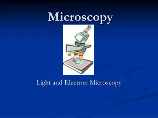
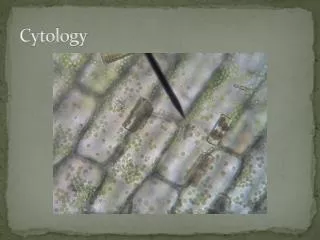
Unit 5. Microscopy & Cytology. Microscopy: Introduction. The unaided eye can distinguish objects more than 0.1 mm in diameter. Since most cells are between 0.1-0.01 mm in diameter, scientists use microscopes to view very small objects such as cells.

E N D
Microscopy: Introduction • The unaided eye can distinguish objects more than 0.1 mm in diameter. Since most cells are between 0.1-0.01 mm in diameter, scientists use microscopes to view very small objects such as cells. • Light microscopes bend visible light using optical devices such as lenses and prisms to magnify the apparent image size. • We will discuss the compound light microscope , you should learn all the parts and their functions, and know the proper care and use of one.
Parts of the Microscope Ocular lens Nose piece Objectives Stage Mechanical stage Condenser Light source Head Arm Coarse knob Fine knob On/off switch Base
Ocular Objectives diaphragm Stage control knobs Condenser Light source
Principles of microscopy • 1. Magnification • Ocular (10x) x objective (40x) = 400x • 2. Parfocal and parcentral imaging • Parfocal: image remains in focus when switching lenses. • Parcentral: image remains in the center of field of view when lenses are changed. Field of view 40x
3. Depth of focus • Is the vertical distance between the lens and the object. Thickness of the specimen decreases as magnification increases. Top A 3-colored thread slide was focused. Note that one thread is in focus and the others are not. The clearly focused thread lies on top of the mount. Middle Bottom 400x
4. Image orientation • The image seen will be real, inverted, and magnified by the objective. • Notice the letter “e” is upside down in the slide. • When viewed through the microscope, it is right side up.
5. Resolving power: the degree at which two adjacent points on a specimen are seen as separate detailed images. • Difference between blurry and sharp images. • 6. Contrast: how well the details of a specimen stand out against a background. Stains and lighting are used to increase contrast to see detail. Protista Volvox seen through the microscope
Cytology: Introduction • In Greek the term for “cell” is “cyto” ; therefore, cytology is the study of cells and how they function. Types of cells • There are two fundamental kinds of cells: prokaryotic and eukaryotic. • Prokaryotes: means “before the nucleus”. Lacks nuclear membrane. • Example: bacteria This is a Bacillus type bacteria • Eukaryotes: “eu” means “real” or “true nucleus”. Eukaryotes can be uni- or multicellular organisms. • Example: plans, animals, protists, fungi. This picture is a dinoflagelate, of the group Protista.
Eukaryotes: Plants • Elodea wet mount • Identify: cell wall, chloroplast, plasma membrane, cytoplasm
cell wall plasma membrane chloroplast cytoplasm
Onion wet mount • This is the membrane between the thick layers of the onion. • Identify: plasma membrane, cell wall, nucleus, nucleolus, cytoplasm.
Animals • Human cheek cells wet mount • Identify: plasma membrane, cytoplasm, nucleus.
plasma membrane nucleus cytoplasm End