Download
1 / 57
600 likes | 1.07k Views
Bowel Elimination. Function- excrete/eliminate waste products of digestion. Maintaining normal bowel elimination is essential to health and efficient body functions. GI System & Functions. Mouth: mostly mechanical digestion ( mastication) Pharynx, Esophagus : passageway for food

E N D
Function- excrete/eliminate waste products of digestion. • Maintaining normal bowel elimination is essential to health and efficient body functions
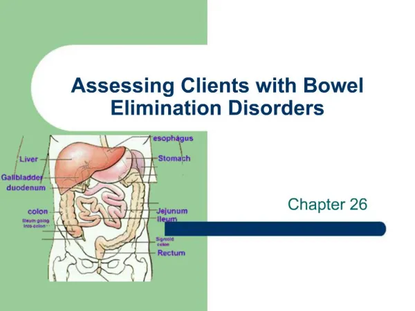
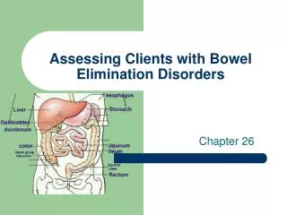
GI System & Functions • Mouth: mostly mechanical digestion (mastication) • Pharynx, Esophagus: passageway for food (from mouth to stomach) • Stomach: bolus is mixed with gastric juice for digestion (liquid, mucus and enzymes) chyme
Liver: - Secretes bile - Processes nutrients - Remove wastes from the body (including old RBCs) - Detoxify - Secretes hormones Pancreas – Secretes enzymes & hormones Gallbladder – Stores bile
Duodenum - Receive juices from pancreas, liver and its own wall • Jejunum-Ileum – Absorption of nutrients • Colon - Reabsorb water from food and digestive juices • Rectum – Storage of feces • Anus - Expels feces and flatus from the rectum
Component of Fecal Matter 1. Water 75%. 2. Solid 25%. • Solidified components of the digestive juices ,undigested fibers (e.gcelluose) which is insoluble ,acts as bowl irritant ,draws water out into lumen ,cleans out lower GIT and is correlated with lower risk of colon cancer. • Dead bacteria (20%). • Fat (10-20%). • Inorganic material (10-20%). • Undigested protein(2-3%).
Properties; • Colour; Normal feces has a dark brown colour, (bilirubin in the presence of bacteria will get oxidized to urobilin which gives stool its typical colour) - Odor; The odor of feces is affected significantly by the type of food ingested and the bacterial flora of the individual (of the main order contributors H2S and mercaptens. - Consistency; Normal feces is solid to semi-solid depending on diet. - 80 - 170 g/day.
Defecation • Rectum usually empty • Mass movement forces fecal matter in • Distention of rectal wall triggers defecation reflex • Stretch receptors in rectal walls stimulate a series of local peristaltic contractions in colon and rectum • Moves feces towards anus • Parasympathetic neurons in sacral region activated by stretch receptors • Stimulate increased peristalsis throughout large intestine • Internal anal sphincter • Must relax so feces can move into anus • External anal sphincter clamps shut • Therefore release is conscious
Factors Affecting Bowel Elimination • Age • Infection • Diet • Fluid Intake • Physical Activity • Psychological factors (Stress …) • Personal Habits/Daily Routine • Position during Defecation • Pain
Pregnancy • Diagnostic GI tests • Surgery and Anesthesia • Medications
Common problems associated with elimination of faeces • Constipation • Fecal Impaction • Diarrhea • Incontinence • Flatulence • Hemorrhoids
Constipation • More of a symptom than a disorder • Straining & pain on defecation is associated symptoms • Can be significant heath hazard (increase ICP, IOP, reopen surgical wounds, cause trauma, cardiac arrhythmias)
NURSING INTERVENTIONS • 1. Assist physician in treating the underlying cause of constipation • 2. Encourage to eat HIGH fiber diet to increase the bulk • 3. Increase fluid intake • 4. Administer prescribed laxatives, stool softeners • 5. Assist in relieving stress
Impaction • Results from unrelieved constipation • Collection of hardened feces wedged into rectum • Can extend up to sigmoid colon • Most at risk: depilated, confused, unconscious (all are at risk for dehydration) • When a continuous ooze of diarrheal stool develops, impaction should be suspected • Associated S/S: Loss of appetite, abdominal distention, cramping, rectal pain
Diarrhea • Increase in number of stools & the passage of liquid, unformed stool (more than 3times/day) • Symptom of disorders affecting digestion, absorption, & secretion of GI tract • Intestinal contents pass through small & large intestines too quickly to allow for usual absorption of water & nutrients
Irritation can result in increased mucus secretion, feces become too watery, unable to control defecation • Excess loss of colonic fluid can result in acid-base imbalances or fluid/electrolyte imbalances • Can also result in skin breakdown
Conditions that cause Diarrhea • Emotional Stress • Intestinal Infection • Food Allergies • Food Intolerance • Tube Feedings (Enteral) • Medications • Laxatives • Colon Disease • Surgery
Medical Management • Treatment of underlying cause • Controlling symptoms & preventing complications • Antibiotics & anti nflammatoryagents • Antidiarrheal & antispasmodic agents • Nursing Management • Assessment & pattern of diarrhea • Bed rest & monitoring of fluid status • Serum electrolytes (K) • Perenial care
Incontinence Inability to control passage of feces and gas from the anus • Caused by conditions that create frequent, loose, large volume, watery stools or conditions that impair sphincter control or function
Medical Management • Bowel training program • Surgical reconstruction • Sphincter repair • Fecal diversion • Nursing Management • Assessment & Health History • Bowel Training program • Maintain skin integrity • Assist patient & family to cope with illness
Flatulence • Gas accumulation in the lumen of intestines • Bowel wall stretches and distends • Common cause of abdominal fullness, pain, & cramping • Gas escapes through mouth (belching), or anus (flatus)
Hemorrhoids • Dilated, engorged veins in the lining of the rectum • External (Clearly visible) or Internal • Caused by straining, pregnancy, CHF, chronic liver disease
NURSING INTERVENTIONS • 1. Advise patient to apply cold packs to the anal/rectal area followed by a SITZ bath • 2. Encourage HIGH-fiber diet and fluids • 3. Administer stool softener as prescribed Post-operative care for hemorrhoidectomy • 1. Position: Prone or Side-lying • 2. Maintain dressing over the surgical site • 3. Monitor for bleeding • 4. Administer analgesics and stool softeners • 5. Advise the use of SITZ bath 3-4 times a day
Nursing interventions to maintain normal bowel elimination pattern • Routine- Establish regular pattern of elimination at regular times. Pt. needs10-15min (uninterrupted time). If urge to defecate is constantly ignored the defecation reflex will be lost, causing feces to remain longer in intestine, increased water absorption, making feces hard and difficult to pass. Use communication skills to discuss bowel patterns. • Positioning- Comfortable position needed. Squatting position common. Assess need for elevated toilet, commode.
Privacy- considered a very private act. Use BR if possible, pull drapes close doors. • Comfort- provide quiet, comfortable as possible place. • Activity- needed to promote GI activity and maintain reg. frequency. Teaching related to inactivity and constipation. Exercises for immobile client. Exercises to strengthen abd. and perineal muscles used for defecation.T & P ROM • Diet/Fluids - High fiber foods, 2000cc fluids/day
High fiber foods: • Legumes (beans) • Cereals • Whole grains • Raw Fruits • Vegetables Laxative effect foods: • Spicy & greasy • Bran/Chocolate • Coffee/Alcohol • Raw fruits & vegetables • Diet
Assessment of Bowel Function • Nursing History • Physical Assessment • Fecal Characteristics • Laboratory/ Diagnostic Tests • Fecal specimens • Guaiac test • Diagnostic tests • Direct visualization • Indirect visualization
Nursing care of the individual with Digestive System Disorders
GASTRITIS/GASTROENTERITIS • Acute gastritis is the irritation and inflammation of the stomach's mucous lining. • Gastritis may be caused by a chemical, thermal, or bacterial insult. • Eg: Drugs such as alcohol, aspirin, and chemotherapeutic agents. • Hot, spicy, rough, or contaminated foods . • Management involves symptomatic treatment measures after removal of the causative agent.
Gastroenteritis, or inflammation of the stomach and intestines, is generally caused by bacteria and viruses. • Other causes include parasites, food allergens, drug reactions to antibiotics, and ingestion of toxic plants. • Treatment is the same as for gastritis, with the addition of anti-microbial drugs for severe cases. • S&S: • Pain, cramping, belching, nausea, and vomiting. Severe cases may include hematemesis. • Diarrhea may occur with gastroenteritis.
. Nursing implications • (1) Stop all P.O. intakes until symptoms subside. • (2) Assess the patient's symptoms and administer the prescribed symptomatic relief medications such as antacids and antiemetics. • (3) Monitor intake and output closely. • Excessive vomiting or diarrhea may result in severe electrolyte depletion that will require replacement therapy. • (4) Administer and monitor IV therapy when ordered to replace lost fluids. • (5) Weigh daily to monitor weight loss. • (6) Encourage the prescribed diet to maintain nutrition.
GASTROINTESTINAL ULCERS • A gastrointestinal ulcer is a break in the continuity of the mucous lining. Ulcers may occur in any part of the GI tract that comes in contact with the gastric juices. (hydrochloric acid and pepsin secretion) • Ulcers commonly occur in the lower esophagus, the stomach, and the duodenum. • Other factors implicated in the development of ulcers. • (1) Emotional stress. • (2) Prolonged physical stress associated with trauma, surgery, burns, and so forth. • (3) Hereditary factors. • (4) Certain drugs and medications. Eg: alcohol, caffeine, aspirin, corticosteroids, and chemotherapeutic agents.
The primary symptom of ulcers is pain. (burning, cramping, aching, or gnawing pain in the stomach area between the xiphoid process and the umbilicus.) • The severity of the pain is generally an indication of the extent of the ulceration. • Pain is normally localized, the patient being able to indicate the area of the pain by pointing one finger. Radiating pain indicates a severe or perforated (ruptured) ulcer.
Nursing implications: • The first objective is to promote gastric rest. • The second objective is prevention of further ulceration. • (1) Encourage physical and emotional rest by using relaxation techniques and prescribed medications (such as sedatives and tranquilizers) to reduce anxiety, restlessness, and insomnia. • (2) Practice prophylaxis (prevention) by use of antacids. Avoidance of irritants such as aspirin, alcohol, caffeine, and spicy foods. • (3) Dietary management aids in control of pain and prevention of ulcers. Meals should be frequent, regular, and small to moderate in size. Foods not well tolerated should be eliminated. Daily intake should be of sufficient caloric and nutritive value to maintain health. (4) When ulceration is in the acute stage, diet should be modified to consist of bland, low-fiber, non-gas-producing foods. Foods that are mechanically, chemically, and thermally nonirritating to the stomach.
Observe for signs and symptoms such as nausea, vomiting, blood in emesis or stool, abdominal rigidity, or abdominal pain. These symptoms may indicate the presence of bleeding, rupture, or obstruction at the ulcer site.
APPENDICITIS • Appendicitis is the inflammation of the vermiform appendix. The appendix fills with food and empties regularly. Because its lumen is quite small, it empties irregularly and is prone to obstruction. The obstruction sets off an inflammatory process that may lead to infection, necrosis, and perforation. • b. Signs and Symptoms. • (1) Generalized abdominal pain that localizes in the right lower quadrant. • (2) Anorexia. • (3) Nausea and vomiting. • (4) Abdominal rigidity or guarding. • (5) Rebound tenderness. • (6) Fever. • (7) Elevated white blood cell count.
Nursing Implications. • (1) Administer IV fluids as ordered to maintain hydration. • (2) Keep the patient NPO until symptoms subside and/or surgery is ruled out. • (3) Position the patient in Fowler's or semi-Fowler's position. This position relaxes the abdominal muscles and reduces pain. • (4) Never apply heat to the abdomen, as this may cause the appendix to rupture. • (5) Analgesics are normally withheld since they mask symptoms.
Treatment. • Treatment of choice is surgical removal of the appendix, especially if rupture is suspected or imminent. • (1) If the appendix can be removed before it ruptures, the post-op course is generally uncomplicated. The wound is closed and the patient is usually discharged within a week. (2) If rupture has occurred, the wound is often left open to drain. The patient must be observed for signs and symptoms of obstruction, peritonitis, hemorrhage, or abscess.
PERITONITIS • The peritoneum is the serous membrane that lines the abdominal cavity and covers the visceral organs. Peritonitis is inflammation of the peritoneum. Inflammation may be generalized throughout the peritoneum, affecting the visceral and parietal surfaces of the abdominal cavity, or may be localized in one area as an abscess. • Peritonitis occurs as a result of leakage of contents from an abdominal organ into the abdominal cavity. • Results from perforation of the GI tract, allowing bacterial contamination of the peritoneum. • Result of chemical irritation, and subsequent infection, caused by rupture of an organ. (For example, the ovaries, spleen, or urinary bladder.)
Signs and symptoms. • (1) Diffuse pain that eventually localizes in the area of the underlying process. • (2) Abdominal tenderness. • (3) Abdominal muscle rigidity. • (4) Nausea and vomiting. • (5) Paralytic ileus. • (6) Fever. • (7) Rapid pulse rate. • (8) Elevated WBC.
Nursing implications. • (1) Observe for signs of hypovolemia and shock. These conditions may result from loss of fluids and electrolytes into the abdominal cavity. • (2) Strictly monitor I&O and vital signs. • (3) Observe safety precautions, since fever and pain may cause the patient to become disoriented. • (4) Administer prescribed medications and intravenous fluid replacement.
INTESTINAL OBSTRUCTION • Intestinal obstruction is defined as any hindrance to the passage of intestinal contents through the small and/or large bowel. • Obstruction may be partial or complete. Severity depends upon the area of bowel affected, the degree of blockage, and the degree of vascular impairment. • Intestinal obstruction is divided into two basic categories: mechanical and non-mechanical.
(1). Mechanical obstruction results from obstruction within the lumen of the intestine or mural obstruction from pressure on the walls of the intestines. Causes include: (a) Foreign bodies such as fruit pits, parasitic worms, or gallstones (b) Volvulus (c) Intussusception. (d) Hernia. (e) Cancer. (f) Adhesions. (g) Strictures.
(2) Non-mechanical obstruction is the result of physiological disturbances. Causes include: • (a) Electrolyte imbalances. • (b) Neurogenic disorders (such as spinal cord lesions). (c) Paralytic (adynamic) ileus, developing as a result of abdominal surgery, trauma, or infection.
Signs and symptoms of large bowel obstruction. • (1) Symptoms of large bowel obstruction differ from those of small bowel obstruction because the colon is able to absorb its fluid contents and distend well beyond normal size. • (2) Constipation may be the only symptom for several days. • (3) Eventually, the distended colon loops will be visible on the abdomen. • (4) Nausea and cramps, abdominal pain will occur. • (5) Vomiting is absent at first, but when obstruction becomes complete, fecal vomiting will occur. • (6) If the obstruction is only a partial one, any of the above symptoms may occur in a less severe form. Additionally, liquid stool may leak around the obstruction.
Nursing implications. • (1) Abdominal girths should be measured daily. • (a) Use the same measuring tape each time. • (b) Place the patient in the same position each time. • (c) Ensure that the tape measure is placed in the same position each time. This can be done by drawing small tic marks on the patient's abdomen to indicate position for the tape. • (d) Measure the patient at the same time each day. • (2) Note the color and character of all vomitus. Test for the presence of occult blood. • (3) Any stool passed should be tested for the presence of occult blood. • (4) Monitor vital signs closely. Elevations of temperature and pulse may indicate infection or necrosis. • (5) Monitor I&O closely. Fluid and electrolyte losses must be replaced.
DIVERTICULAR DISEASE • Diverticula are bulging dilatations or "out-pouchings" of the gastrointestinal walls. Common sites are the sigmoid colon, duodenum, and the distal ileum. Occur anywhere along the GI tract, from the esophagus to the anus. • Diverticulosis. The presence of asymptomatic diverticula is called diverticulosis. • Diverticulosis pain that is relieved by defecation or flatulence. • Constipation or diarrhea may also occur. • Diverticulosis generally requires no treatment other than dietary modification to prevent irritation of the bowel.
c. Diverticulitis- inflamed or infected diverticula. Food and bacteria lodge and harden in the diverticular sac. Inflammation results, followed by infection. Complications include abscess, obstruction, perforation, peritonitis, and hemorrhage. • (1) Symptoms • Low grade fever, nausea, gas, abdominal pain, and abdominal rigidity. • (2) Treatment • Mild cases of diverticulitis includes antibiotics, antispasmodics, stool softeners, and liquid diet. • Severe cases of diverticulitis, or cases that involve perforation, obstruction, fistula, or peritonitis may require surgical intervention. Colon resection may be necessary to remove the diseased portion of the bowel. A temporary or permanent colostomy may be indicated.
Nursing Implications. • (1) Reinforce patient education regarding dietary modification. Increased roughage in the diet may prevent intestinal contents from lodging in the diverticula. Roughage includes grains, fruits, vegetables, and fiber. • (2) When symptoms occur, the patient should immediately alter his diet to one that is bland and non irritating. • (3) Diet should include adequate fluid intake to avoid constipation. Constipation encourages inflammation of the bowel. • (4) Vital signs and I&O should be monitored closely. • (5) Observe stools for color and consistency. • (6) If surgery becomes necessary, observe routine preoperative and postoperative nursing care procedures.
Liver Cirrhosis • A chronic, progressive disease characterized by a diffuse damage to the hepatic cells • The liver heals with scarring, fibrosis and nodular regeneration ETIOLOGY: Post-infection, Alcohol, Cardiac diseases, Schisostoma, Biliary obstruction