Download
1 / 37
390 likes | 640 Views
Venous Insufficiency: Nuts and Bolts. Harry Ma MD, PhD Assistant Professor of Surgery University of Oklahoma Tulsa, Oklahoma. Department of Surgery. Disclosures. None. Outline and Objectives. Defining chronic venous disease Clinical manifestations Diagnostic evaluation Treatment

E N D
Venous Insufficiency: Nuts and Bolts Harry Ma MD, PhD Assistant Professor of Surgery University of Oklahoma Tulsa, Oklahoma Department of Surgery
Disclosures • None
Outline and Objectives • Defining chronic venous disease • Clinical manifestations • Diagnostic evaluation • Treatment • Non-operative • Operative
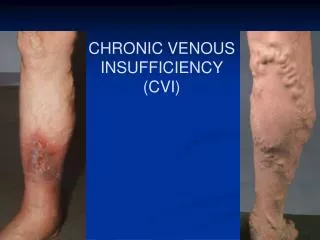
Spectrum of Disease • Spider veins (telangiectases) • Reticular veins • Varicose veins • Edema • Chronic skin changes • Ulcers
Impact of CVD • Most common form of vascular disorder • Chronic Venous Disease: 25 million people in US • Health care cost: $1 to 3 Billion dollars annually • Indirect cost: ~2 million work days lost annually
Definitions • Telangiectasias - are a confluence of dilated intradermal venules less than one millimeter in diameter. • Reticular veins - are dilated bluish subdermal veins, one to three millimeters in diameter. Usually tortuous. • Varicose veins - are subcutaneous dilated veins three millimeters or greater in size. They may involve the saphenous veins, saphenous tributaries, or nonsaphenous superficial leg veins.
Definitions of Skin Changes • Lipodermatosclerosis: localized chronic inflammation and fibrosis of the skin and subcutaneous tissue • Atrophie blanche: localized, often circular whitis and atrophic skin areas surrounded by dilated capillary spots and hyperpigmentation • Venous Ulcer: full thickness skin defect
CEAP classification • Clinical • Etiology • Anatomy • Pathophysiology
Clinical Classification • C0: no visible or palpable signs of venous disease. • C1: telangiectasies or reticular veins. • C2: varicose veins. • C3: edema. • C4a: pigmentation and eczema. • C4b: lipodermatosclerosis and atrophie blanche. • C5: healed venous ulcer. • C6: active venous ulcer.
Etiologic Classification • Ec: congenital (<5%) • Ep: primary(65-80%) • Es: secondary (postthrombotic 15-28%).
Valvular Dysfunction • Primary valvular dysfunction: weakness in leaflets or vessel wall • Secondary to previous DVT or phlebitis • Increases retrograde flow • Results in reflux • Increased hydrostatic pressure • Not just deep or superficial valves but perforators
Diagnostic Evaluation • Venous Duplex (New Gold Standard) • Phlebography or Venography (Old Gold Standard) • Air Plethysmography • Photophlethysmography • Venous pressure (hemodynamic gold standard)
Venous Duplex • Can rule out thrombosis and obstruction • Quantify reflux in veins • Visualize anatomy
Venous Duplex • Vast majority have superficial incompetence only. • Sensitivity 95 % for identifying the competence of the saphenofemoral and saphenopopliteal junctions. • Less sensitive for identifying incompetent perforators (40 to 60 percent)
Risk Factors for varicose veins • Morbid obesity • Advanced age • Sex/Hormonal changes • Family history/Genetics • History of DVT or phlebitis • Occupational risks
Clinical Manifestations • Veins are prone to thrombophlebitis • Common symptoms: • Pain • Swelling • Ulcerations • Skin changes • Cramping • Fatigue
Clinical Manifestations • Itching • Burning • Pain after standing • Relieved with leg elevation • Vague pain • Complications: • Ulceration • Bleeding • Skin changes
Patient Assessment • History • History of symptoms and onset • History of venous complications • Desire for treatment • Comorbidities • Rule out secondary cause including DVT and HEART Failure • Examination • Patient in general • Pedal pulses • Groins • Veins • Studies • Venous Duplex
Non-operative management • Leg elevation • Compression therapy • Treatment of ulcer: local wound care • NOT DIURETICS
Non-operative management • LEG ELEVATION – heart level for 30 minutes 3-4 times daily improves micro-circulation reduces edema, and promotes healing of venous ulcers. • EXERCISE – daily walking and simple ankle flexion exercises.
Compression Therapy • Compression bandages • Compression Stockings • Intermittent Pneumatic Compression • Contraindications • Active infection • Significant arterial occlusive disease • ABI < 0.6-0.8
Compression Bandages Elastic In-elastic • Types: ACE wrap • Easier to use • Higher pressures at rest • Can cause pressure ulcers • Minimal increase in pressure when ambulatory • Types: Profore multi-layer wraps • High stiffness • Exert about 40mmHg • Can lose pressure quickly due to limb volume reduction • Difficult to apply • Better for ambulatory patients
Benefits of Compression • Increased ulcer healing • 30-40mmHg compression resulted in 93% ulcer healing rate by 6 months • Prevention of recurrence with compliance: 29% recurrence at 5 years • Disadvantages: Compliance
Operative Management • Ligation and Stripping • Ablative therapy • Radiofrequency • Laser • Sclerotherapy
Ligation and Stripping • Traditional surgical approach • Reduced recurrence rates compared to high ligation alone
Ablation • Laser • Use a bare tipped optical fiber which applies laser light energy to the vein. • Therapy based on photothermolysis (light induced thermal damage). • Laser light heats the target tissue inducing thermal injury • Wavelength of light is chosen based on the target structure's chromophore • Radiofrequency • A high frequency alternating current resulting in energy that heats the adjacent vein walls to the probe which alters the protein structure of the vein effecting its closure
Complications of ablation • Nerve injury <2% • Skin injury <1% • Failure of closure <5% • Thrombotic complications <1%
Which is Better? • Equivalent therapy • Similar outcomes • Success rates >95% for both (N=159) • Improvement in QoL surveys and VVQ surveys • Cochrane review in 2014 • 13 randomized trials included • 3081 patients total • Complication rates equivalent • Efficacy: equivalent for laser, RFA, foam sclerotherapy and ligation and stripping
Sclerosing Agents • Hypertonic Saline • Chromated glycerin • Nonchromated glycerin • Monoethanolamineoleate • Sotradecol (STS) • Polidocanol • Foam
Mechanism of action and use • Endothelial damage • Thrombosis • Resultant sclerosis and closure of the vein • For telangiectasias • Reticular veins • Perforator veins • More recently incompetent GSV
Ultrasound Guided Foam Sclerotherapy • First reported in 1995 • Mixed with air or CO2 • Bubble size of 100 µm or less • Ration of sclerosant to air or CO2 is usually 1:4
Limitations and complications • Large veins • Phlebitis 3-8% • Embolus (CVA) • Visual disturbance • DVT • Failure or recurrence • Anaphylaxis
Short term outcomes • 1 year follow-up equivalent QoL improvement • 72% success rate (vs 89% for EVLA) • Two year follow-up in patients with ulcers • 85% healing rate
Long term outcomes • 5-8 year follow-up of 285 patients • 89% had improvement in QoL survey and AVSS • 15% required repeat treatment
References • André P, Hartwell D, Hrachovinová I, Saffaripour S, Wagner DD. Pro-coagulant state resulting from high levels of soluble P-selectin in blood. ProcNatlAcadSci U S A 2000;97:13835-40. PMID: 11095738Beebe-Dimmer JL, Pfeifer JR, Engle JS, Schottenfeld D. The Epidemiology of Chronic Venous Insufficiency and Varicose Veins. Ann Epidemiol 2005;15:175-84. PMID: 15723761 • Bradbury A, Evans C, Allan P, Lee A, Ruckley V, Fowkes FG. What are the symptoms of varicose veins? Edinburgh vein study cross sectional population survey. BMJ 1999;318:353-6. PMID: 9933194. • Bradbury A, Evans CJ, Allan P, Lee AJ, Ruckley CV, Fowkes FG. The relationship between lower limb symptoms and superficial and deep venous reflux on duplex ultrasonography: The Edinburgh Vein Study. J VascSurg 2000;32:921-31. PMID: 11054224 • Browse NL. The diagnosis and management of primary lymphedema. J VascSurg 1986;3:181-4. PMID: 3510325 • Burnand KG, Whimster I, Naidoo A, Browse NL. Pericapillary fibrin in the ulcer-bearing skin of the leg: the cause of lipodermatosclerosis and venous ulceration. BMJ (Clin Res Ed) 1982;16;285:1071-2. PMID: 6812751 • Cardinal M, Eisenbud DE, Phillips T, Harding K. Early healing rates and wound area measurements are reliable predictors of later complete wound closure. Wound Repair Regen 2008;16:19-22. PMID: 18211575
References • Carpentier PH, Maricq HR, Biro C, Ponçot-Makinen CO, Franco A. Prevalence, risk factors, and clinical patterns of chronic venous disorders of lower limbs: a population-based study in France. J VascSurg 2004;40:650-9. PMID: 15472591 • Carpentier PH, Poulain C, Fabry R, Chleir F, Guias B, Bettarel-Binon C; Venous Working Group of the SociétéFrançaise de MédecineVasculaire. Ascribing leg symptoms to chronic venous disorders: the construction of a diagnostic score. J VascSurg 2007;46:991-6. PMID: 17980285 • Christopoulos D, Nicolaides AN, Szendro G. Venous reflux: Quantitation and correlation with the clinical severity of chronic venous disease. Br J Surg 1988;75:352-6. PMID: 3359149 • Comerota AJ, The ATTRACT Trial: Rationale for early intervention for iliofemoral DVT. PerspectVascSurgEndovasTher 2009; 21(4): 221-4. Epub 2010 Jan 3. PMID: 20047905 • Comerota AJ, Throm RC, Mathias SD, Haughton S, Mewissen M. Catheter-directed thrombolysis for iliofemoral deep venous thrombosis improves health-related quality of life. J VascSurg 2000;32:130-7. PMID: 10876214 • Cornu-Thenard A, Boivin P, Baud JM, De Vincenzi I, Carpentier PH. Importance of the familial factor in varicose disease. Clinical study of 134 families. J DermatolSurgOncol 1994;20:318-26. PMID: 8176043 • Corriere MA, Suave KJ, Ayerdi J, Craven BL, Stafford JM, Geary RL, Edwards MS: Vena cava filters and inferior vena cava thrombosis. J VascSurg 2007;45:789-94. PMID: 17398389