Download
1 / 1
10 likes | 212 Views
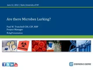
Lurking Oral Cavity Microbes Teddy Annang, Lacey Ebert, Hannah Goldammer, Belarmino Hafermann. Introduction

E N D
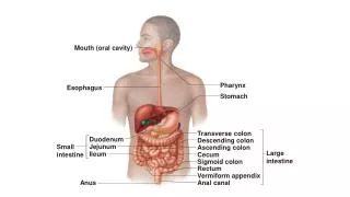
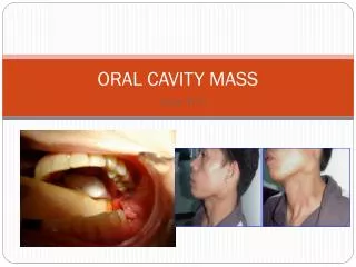
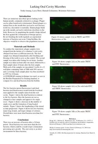
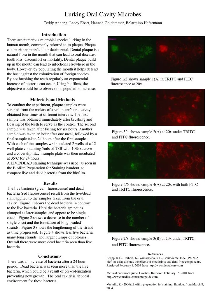
Lurking Oral Cavity Microbes Teddy Annang, Lacey Ebert, Hannah Goldammer, Belarmino Hafermann Introduction There are numerous microbial species lurking in the human mouth, commonly referred to as plaque. Plaque can be either beneficial or detrimental. Dental plaque is a natural flora in the mouth that can lead to oral diseases, tooth loss, discomfort or mortality. Dental plaque build up in the mouth can lead to infections elsewhere in the body. However, by populating the mouth it helps defend the host against the colonization of foreign species. By not brushing the teeth regularly an exponential increase of bacteria can occur. Using biofilms, the objective would be to observe this population increase. Figure 1/2 shows sample 1(A) in TRITC and FITC fluorescence at 20x. Materials and Methods To conduct the experiment, plaque samples were scraped from the molars of a volunteer’s oral cavity, obtained four times at different intervals. The first sample was obtained immediately after brushing and flossing of the teeth to serve as the control. The second sample was taken after fasting for six hours. Another sample was taken an hour after one meal, followed by a final sample taken 24 hours after the first sample. With each of the samples we inoculated 2 wells of a 12 well plate containing 5mls of TSB with 10% sucrose and a coverslip. Each sample plate was then incubated at 35ºC for 24 hours. A LIVE/DEAD staining technique was used, as seen in the Biofilm Preparation for Staining handout, to compare live and dead bacteria from the biofilm. Figure 3/4 shows sample 2(A) at 20x under TRITC and FITC fluorescence. Results The live bacteria (green fluorescence) and dead bacteria (red fluorescence) result from the live/dead stain applied to the samples taken from the oral cavity. Figure 1 shows the dead bacteria in contrast to the live bacteria. Here the bacteria are not as clumped as later samples and appear to be single cocci. Figure 2 shows a decrease in the number of single cocci and the formation of long beaded strands. Figure 3 shows the lengthening of the strand as time progressed. Figure 4 shows less live bacteria, many long strands, and larger clumps of colonies. Overall there were more dead bacteria seen than live bacteria. Figure 5/6 shows sample 4(A) at 20x with both FITC and TRITC fluorescence. Figure 7/8 shows sample 3(B) at 20x under TRITC and FITC fluorescence. References Kropp, K.L., Herbort, K., Wimalasena, R.L., Goulbourne, E.A. (1997). A biofilm assay at study the effects of mouthrinse and dentifrice components. Retrieved February 4, 2004 from http://www.dentalcare.com Medical consumer guide. Cavities. Retrieved February 16, 2004 from http://www.medicalcomsumerguide.com Ventullo, R. (2004). Biofilm preparation for staining. Handout from March 8, 2004. Conclusions There was an increase of bacteria after a 24 hour period. Dead bacteria was seen more than the live bacteria, which could be a result of pre-colonization preventing new growth. The oral cavity is an ideal environment for these bacteria.