Download
1 / 63
630 likes | 747 Views
Is There a Role for Nonivasive Imaging Strategies For Risk Stratification in ACS?. George A. Beller MD No Disclosures. Clinical History.

E N D
Is There a Role for Nonivasive Imaging Strategies For Risk Stratification in ACS? George A. Beller MD No Disclosures
Clinical History Mr. H is a 63 year-old male with a past history of hypertension, hyperlipidemia, and uncontrolled Type 2 diabetes mellitus who presented with a 3-day history of progressive shortness of breath and lower extremity edema. He recalled having some vague substernal chest discomfort lasting approximately 2-3 hours associated with the onset of his SOB three days previously.
PMH: Newly-diagnosed CHF Type 2 DM Hypertension Dyslipidemia Meds: Verapamil, Lisinopril, Lasix, Pravastatin, Indomethacin, Metformin Social Hx: Lives with his wife No known ETOH, tobacco, or drugs PE: 114/65, 82, 18 Increased JVP to 3cm above clavicle Lungs- bibasilar crackles RRR, no MRG 2+ LE edema, 2+ pulses Labs: K 4.1, Cr 1.5, Gluc 379 BNP 1429 Troponins 2.4, 1.8, 1.2 Hct 51.3 CXR: Mild pulmonary venous congestion
Hospital Admission • He was diagnosed with CHF secondary to an MI which occurred 3 days previously. • LVEF was 15-20% by echo.
Admission • He was successfully diuresed, the verapamil discontinued and started on metoprolol • He then underwent cardiac cath showing the following: proximal LAD: 100% mid LCx: 70% PDA: 80%
Question Which of the following would be the next step in management? Refer directly to cardiac cath for PCI of the total LAD occlusion 2. Refer the patient directly for CABG. 3. Perform a noninvasive imaging study of ischemia/viability. 4. Continue with medical therapy without revascularization
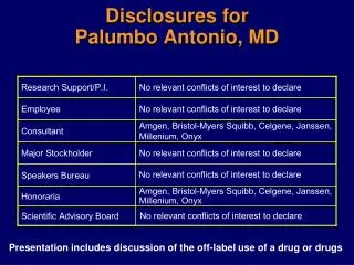
Subsequent Evaluation He underwent adenosine stress/rest SPECT sestamibi myocardial perfusion imaging to assess for viability and ischemia.
Sestamibi Stress/ Rest Images Quantitative Perfusion
Sestamibi Rest Gated Images . Quantitative Function
Question Which of the following would be the next step in management? The patient has poor viability and should be treated medically. 2. The patient should have a cardiac MRI study to assess extent of scar. 3. The patient has preserved viability and would have a better long term outcome with revascularization.
Subsequent Management It was decided to proceed with coronary revascularization based on the finding of moderately preserved viability in the distribution of all 3 coronary territories. Four weeks later he returned for a follow-up adenosine stress sestamibi perfusion imaging study
Sestamibi Stress / Rest Images G.A. Quantitative Perfusion- post revascularization
Sestamibi Stress / Rest Images G.A. Quantitative Function- post revascularization
The Message The patient appeared too late after his MI for primary PCI of his totally occluded LAD to attain myocardial salvage with RP He had multivessel CAD with severe LV dysfunction but had preserved viability in all 3 coronary territories. Such patients with multivessel CAD and severe LV dysfunction benefit from a viability study prior to considering revascularization.
Myocardial Imaging in ACS • Detection of CAD in low-intermediate risk patient presenting with chest pain in ED • Noninvasive assessment of LV function and infarct size for prediction of remodeling • Stress imaging for risk stratification • Determination of functional significance of intermediate stenosis (50%-70%) • Assessment of viability in infarct zone to determine value of late reperfusion
Immediate Evaluation for Possible ACS Class I In pts with suspected ACS in whom IHD is present or suspected, if the follow-up ECG and cardiac biomarker levels are normal, a stress test (exercise or pharmacogic) to provoke ischemia should be performed in the ED, in a CPU, or on an outpatient basis in a timely fashion (within 72hr) as an alternative to inpatient admission. Low risk patients with a negative diagnostic test can be managed as outpatients (Level of Evidence: C)Class I Patients with possible ACS and negative biomarkers who are unable to exercise or who have an abnormal resting ECG should undergo a pharmacologic stress test (Level of Evidence: B). (ACC/AHA UA/NSTEMI Guideline Revision, Circ 2007;116:803-877)
Immediate Evaluation for Possible ACS -2 Class IIa In pts with suspected ACS with a low or intermediate probability of CAD, in whom the follow-up 12-lead ECG and cardiac biomarker measurements are normal, performance of a non-invasive coronary imaging test (i.e. coronary CT angiography) is reasonable as an alternative to stress testing. (Level of Evidence: B)Class I Patients discharged from the ED or CPU should be given specific instructions for activity, medications, additional testing and follow-up with a personal physician. (Level of Evidence: C) (ACC/AHA UA/NSTEMI Guideline Revision, Circ 2007;116:803-877)
Chest Pain And Possible Acute Coronary Syndrome Troponin I; Serial ECGs; Observation Positive Admit Stat ECG, rest MPI or Echocardiogram Recurrent Pain 8-10 hrs Negative Findings ECG Stress Testing, Stress Imaging or CTA Positive Negative Admit Home
Resting Sestamibi Scan in Patient With Chest Pain And Normal Initial ECG SA VLA HLA (Udelson, Heart 2004; 90: v16-v25)
Negative Predictive Value of Resting MPI For Acute MI in The ED
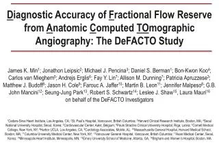
64-Multidetector Row CTA in Patients Without Known CAD (ACCURACY Trial) 99 99 95 100 94 83 83 64 48 % 50 0 Sens Spec PPV NVP Sens Spec PPV NVP Patients (≥ 50% Stenosis) Patients (≥ 70% Stenosis) (Budoff, JACC 2008; 52: 1724-32)
CTA vs Standard of Care (SOC) in Chest Pain (Goldstein JACC 2007; 49: 863-71)
CT Characteristics of Culprit Plaques in 2 Pts. (Cyrus, J Nucl Cardiol 2009; 16: 466-73)
ACS- Free Survival vs. Positive Vessel Remodeling and Low Attenuation Plaques on CTA (Motoyama, JACC 2009: 54: 19-57)
SPECT Imaging After Acute ST Segment Elevation MI • Quantitation of infarct size • Assessment of infarct zone viability • Identify myocardial stunning after reperfusion • Residual infarct zone ischemia in patients not undergoing angiography (stress imaging) • Determination of functional significance of non-infarct stenoses (stress imaging)
Diagnosis, Risk Assessment, Prognosis and Assessment of Therapy After Acute STEMI Indication Test Class • Detection of ischemia and myocardium at risk in thrombolytic pts. without cath. • Infarct size and residual viable myocardium in acute STEMI Stress/ Rest MPI 1 B Rest/ Stress MPI 1 B (ACC/AHA Guidelines, 2003)
Correlation Between Defect Size by Sestamibi SPECT And Myocardial Scar Pathology 70 60 50 40 30 20 10 0 Defect by SPECT (%) Y = 6.60 + 1.03 x r = 0.89, P< 0.001 0 10 20 30 40 50 60 70 Scar by Pathology (%) (Medrano, Circ 1996; 1010-17)
SPECT Infarct Size vs 6 Months Mortality P= 0.006 6 N= 137 N= 129 4 N= 251 6 Month Mortality (%) N= 181 2 N= 424 0 <12 12-19 20-35 36-50 >50 Infarct Size LV (%) (Alamanni, Heart 2004; 90: 1291-98)
99mTc-Sestamibi Infarct Size at Hospital Discharge vs LVESV at One Year (Chareonthaitawee, JACC 1995; 25: 567)
Delayed Hyperenhancement MRI in Acute Transmural MI With No-Reflow (Shan, Circ 2004; 109: 1328-34)
Viability Assessment Predicts Future Ventricular Remodeling After Acute MI And Treatment With Primary Angioplasty Infarct-Zone Viability Variables Yes No End-diastolic Volume 53 14 76 18 End-systolic Volume 22 11 42 17 6 mo. patency rate (%) 98 96 Baseline EF (%) 45 11 44 10 (Bolognese, Circ 1997; 96: 3353-3359)
PCI vs Medical Therapy in OAT Trial Total Events Nonfatal Myocardial Infarction (Hochman, NEJM 2006; 355: 2395)
Risk Stratification Before Discharge: UA/NSTEMI Class I Nononvasive stress testing is recommened in low-risk patients who have been free of ischemia at rest or with low-level activity and of HF for a minimum of 12 to 24 hrs (Level of Evidence:C)Class I Noninvasive stress testing is recommended in patients at intermediate risk who have been free of ischemia at rest or with low-level activity and of HF for a minimum of 12 – 24 h) (Level of Evidence:C) Choice of stress test is based on the resting ECG, ability to perform exercise, local expertise and technologies available; treadmill exercise is useful in patients able to exercise in whom the resting ECG is free of abnormalities (e.g. LVH, BBB, ST ↓) (ACC/AHA UA/NSTEMI Guideline Revision, Circ 2007;116:803-877)
Risk Stratification Before Discharge (Cont’d) Class I An imaging modality should be added in patients with resting ST depression, LVH, BBB, intraventricular conduction defect,preexcitation, or digoxin who are able to exercise. In patients undergoing a low-level exercise test, an imaging modality can add sensitivity (Level of Evidence: B) Class I Pharmacologic stress testing with imaging is recommended when physical limitations (e.g. arthritis, amputation, severe peripheral vascular disease, severe COPD, orgeneral debility) preclude adequate exercise stress. (Level of Evidence: B (ACC/AHA UA/NSTEMI Guideline Revision, Circ 2007;116:803-877)
Cardiac Death (CD) or Nonfatal MI (MI) in Non-ST Elevation ACS Based on NI Testing Negative Positive 25 20 15 % CD/ M 10 5 0 Stress ECG Stress Myocardial Perfusion Imaging (Udelson, Heart 2004; 90: v16-v25)
Events Related to METS in Post-MI Patients Proportion of Death or re-MI 0.20 0.15 0.10 0.05 0.00 < 6 METS 6 -8 METS > 8 METS 0 1 2 3 Years (Valuer, Eur Heart J 2005;26:119-27)
Event Rate in Acute Uncomplicated First MI Patients vs DP Sestamibi Findings 18 Extent of Reversibility 16 Low 14 Intermed 12 High Annual Cardiac Death or MI Rate (%) 10 8 6 4 2 0 Low SSS Intermed High SSS SSS SSS = Summed Stress Score Summed Difference Score used for Ischemia Extent (Brown, Circulation,1999)
INSPIRE Study Design Stable pts following Acute MI Adenosine Sestamibi SPECT #1 (N=728) PDS <20% (N=242) PDS >20%, IPDS >10% (N=273) PDS >20%, IPDS <10% (N=213) LVEF <35% (N=68) LVEF >35% (N=205) Coronary Angiography Strategy 1 Strategy 2 Intensive Medical Rx PTCA/CABG + Medical Rx Revascularization and/or Medical Rx Adenosine Sestamibi SPECT #2 - Blinded Analysis Follow-up - 1 year
Perfusion Defect Size vs Events After AMI Cardiac Events (%Patients) Perfusion Defects Size (%LV) (Mahmarian, JACC 2006; 48: 2448-57)
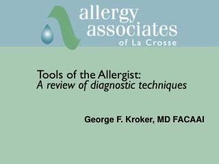
1.0 0.9 0.8 0.7 0.00 0.20 0.40 0.60 0.80 1.00 Risk Based on Ischemic Defect Size Overall Event-Free Survival 0 (n=167) 1-6 (n=345), RR=1.78, p=0.11 Event-Free Survival 7-14 (n=127), RR=2.80, p=0.008 15-20 (n=35), RR=4.75, p=0.001 >20 (n=54), RR=6.15, p<0.0001 Time to Follow-up (Year)
Outcome vs Therapy in INSPIRE Trial of Acute MI (Mahmarian, JACC 2006; 48: 2458-67)
Initial Conservative vs Initial Invasive Strategy Class I An early invasive strategy (diagnostic angiography with intent to perform revascularization) is indicated in initially stabilized UA/NSTEMI patients (without serious comorbidities or contraindications to such procedures) who have an elevated risk for clinical events (Level of Evidence A)Class IIb In initially stabilized patients, an initially conservative strategy (i.e. selective invasive) may be considered as a treatment strategy for UA/NSTEMI patients who have an elevated risk for clinical events including those who are troponin positive. The decision to implement an initial conservative strategy may be made by considering physician and patient preference (LOE: C) (ACC/AHA UA/NSTEMI Guideline Revision, Circ 2007;116:803-877)
Initial Conservative vs Initial Invasive Strategy Class III An early invasive strategy (i.e. diagnostic angiography with intent to perform revascularization) is not recommended in patients with acute chest pain and a low liklihood of an ACS (Level of Evidence: C)Class III An early invasive strategy is not recommended in patients with extensive comorbidities (e.g. liver or pulmonary failure, cancer), in whom the risks of revascularization and comorbid conditions are likely to outweigh the benefits of revascularization (Level of Evidence: C) (ACC/AHA UA/NSTEMI Guideline Revision, Circ 2007;116:803-877)
Long-Term Outcome of Routine Invasive (RI) vs. Selective Invasive (SI) Strategy In Patients With Non-STEMI ACS Pooled analysis from FRISC-II, RITA-3 and ICTUS Over 5 years 14.7% of patients randomized to an RI strategy experienced CV death or MI vs 17.9% in the SI strategy (P=0.002) Differences in CV death not significant (P=0.068) (Fox, JACC 2010; 55:2435-45)
Long-Term Outcome of Routine Invasive (RI) vs Selective Invasive (SI) Strategy In Patients With Non-STEMI ACS (con’t) d. There were 2.0% to 3.8% absolute reductions in CV death or MI in the low- and intermediate-risk groups and an 11.1% absolute risk reduction in the highest-risk patients. e. During 5 years of observation, the majority of patients RI and SI ultimately underwent PCI or CABG (81% vs 60%). (Fox, JACC 2010; 55:2435-45)
Meta-Analysis for CV Death or MI (FRISC-II, RITA-3, ICTUS) Selective Invasive vs. Routine Invasive. Study FRISC-II RITA-3 ICTUS Overall 0.5 0.75 1 1.33 2 Favors Routine Invasive Favors Selective Invasive Hazard Ratio (Fox KAA et al JACC 2010; 55: 2435-45)
Cumulative Risk of CV Death or MI Comparing Routine Invasive vs. Selective Invasive Strategies for ACS (Fox, JACC 2010; 55: 2435-45)
Conclusions 1. Noninvasive imaging for detection of ACS or CAD useful in evaluation of chest pain in the ED for patients with nondiagnostic ECGs and negative biomarkers. 2. Rest imaging after acute MI can estimate infarct size and determine extent of infarct zone viability which have prognostic value.