Download
1 / 30
300 likes | 453 Views
G-Mod Lab Activity Detecting Genetically Modified Foods. Review the use of micropipettes DNA extraction from food samples using gene releaser and temperature cycle Prep PCR components using extracted DNA from #2. 4. Run the PCR program 5. Prepare the samples for loading 6. Load gels

E N D
G-Mod Lab ActivityDetecting Genetically Modified Foods • Review the use of micropipettes • DNA extraction from food samples using gene releaser and temperature cycle • Prep PCR components using extracted DNA from #2
4. Run the PCR program 5. Prepare the samples for loading 6. Load gels 7. Run gels 8. Analyze gels
Micropipetting Set micropipette to proper amount – be sure you are using the correct volume micropipette (.5-10uL, 2-20uL, 20-200uL or 40-200uL) Lock the setting once the desired volume has been located. Obtain a pipette tip from the tip box Depress the push button and stop at the first stop
Micropipetting - continued 5. Place the tip into the sample to be drawn up 6. Release the push button slowly to draw up the sample 7. Position the pipette tip in the proper tube 8. Depress the push button all the way to expel the liquid 9. Remove the micropipette from the tube 10. Release the micropipette push button * Be sure to change tips between each solution
Practice Micropipetting In your group of two, use the two micropipettes at your station to measure the following volumes of solutions. Each person should do each volume. Be sure to change tips between solutions • 100 uL 2. 4 uL
Protocol for DNA extraction from sample using GeneReleaser • Each group of 4 will select one of the food sources • Proceed to the Student Procedures (p. 27 – bottom right of page) and begin with #1, work through number 9 – work through these steps together We will all finish together and begin the temperature cycle to extract DNA – 20 minutes
DNA Extraction using Gene Releaser • 1. Label the top of the sample tube with your initials – clear top tubes • 2. Crumble food sample into powder in sterile lab tissue paper. Transfer a small amount of food into sample tube – approximately one pinky • 3. Add 500 uL of distilled water to the sample and mix using the plastic disposable pestle • 4. Centrifuge for 1 minute to obtain a clear supernatant – Be sure to balance tubes in microcentrifuge
5. If no supernatant is present, add 100 additional uL of distilled water, mix by inverting the tube and pellet undissolved materials by centrifugation for 1 minute. • 6. If supernatant is present, label one of the small clear 200 uL tubes from the kit with your initials • 7. Pipette 50uL of GeneReleaser up and down within the GeneReleaser tube from the student kit to mix the contents thoroughly. Transfer 50uL of GeneReleaser into the newly labeled clear 200uL tube
8. Transfer 10uL of the supernatant from step 5 into the 200uL tube that now contains the 50uL of GeneReleaser. Mix gently with the micropipettor, being careful to leave all the liquid in the tube when you remove the micropipettor • 9. Place the tube in the MyCube Thermal Cycler – record your position on the table at the cycler
GeneReleaser Temperature Cycle • The GeneReleaser Temperature Cycle will run for 20 minutes. This is NOT a polymerase chain reaction process – It is a program of varying temperatures for the extraction of DNA • Once the DNA has been extracted, we will prepare the sample for PCR amplification
PCR – Amplification of DNA • 1. Label the small colored tube found in the student kit with your initials. The 0.5 mL screw cap microcentrifuge tube (purple top) contains the chemicals needed for the PCR amplification of the plant and GMO vector DNA. This PCR master mix is in powdered form and needs to be mixed with water. Add 90uL of distilled water and mix the contents well by inverting the tube 10 times.
2. One partner from each pair should transfer 42uL of the reconstituted PCR mix into their labeled small colored tubes. • 3. Obtain the tube containing the GeneReleaser and extracted DNA. If the GeneReleaser chemical comes into contact with the PCR master mix, it will interfere with the amplification process. To prevent this from occurring, the tube must be centrifuged to pellet any remaining GeneReleaser. Place the clear small tubes containing GeneReleaser and extracted DNA into the emptied distilled water tube. Make sure smaller tubes are capped tightly. Run samples for 2 mintues
4. Once centrifuged, carefully remove the smaller tube from the larger one and transfer 8uL of clear supernatant, containing the DNA, into the colored tube containing the water and PCR mix. Make sure not to transfer any of the GeneReleaser (the white material at the bottom of the tube). • 5. Place the tubes in the MyCube. Record the location of the tubes. • Begin PCR Amplification
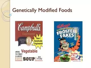
What are genetically modified organisms? (GMOs) • GMOs are organisms in which the genetic material (DNA) has been altered or modified through a method that does not occur in nature – this is often done to introduce a new or altered characteristic to the modified organism
Why develop GMOs? • Modification of agriculturally important crops For example: Bacillus thuringensis produces a chemical that is toxic to some crop-damaging insects, but not to beneficial insects Roundup Ready crops – Agrobacteriumtumifaciens – produces a protein that introduces resistance to the killing effect of Roundup – weeds are still killed and crops are immune to the effects
What are the potential benefits of GM products? • Crops – enhanced taste and quality, reduced maturation time, increased nutrients, yields and stress tolerance, improved resistance to disease, pests and herbicides • Animals –increased resistance, productivity and hardiness, better yields of meat, eggs and milk • Environment – “friendly” bioherbicides and bioinsecticides, conservation of soil, water and energy, better natural waste management
Why is there controversy over GMOs? • Potential human health impacts – allergens, unknowns • Domination of world food production by a few companies • Tampering with nature by mixing genes among species • Labeling is not mandatory in some countries (U.S.)
Why test for the presence of GMOs? • Labeling standards vary : U.S. – any product containing less than 5% GMO may be labeled as GM free Europe – will not accept imports unless they are 0.9% GM or less
What is PCR? • Molecular copy machine – copies a specific region of DNA • 40 cycles: Temperature is increased to 95 C – DNA strands separate – denature Temperature is cooled to between 50-60 C – annealing – primers bind to DNA Temperature is increased to 72 C, activating polymerase, adding nucleotides to the ends of the primers and extending a new copy of DNA strand
Primers used in the PCR reaction: • NPTII gene marker – for GMO detection • 16S plant ribosomal marker – control primer
What has been amplified and what are we looking for? • 1. NPTII – gene that is part of the vector used to create many genetically modified organisms – it is 254 base pairs in length • 2. The second gene is a 16S ribosomal gene associated with plant chloroplast DNA – this serves as a control – because it is present in plants – the amplified portion of the 16S ribosomal gene is 525 base pairs in length
How does electrophoresis work? • Gel electrophoresis
Flash Gel Analysis • 1. Obtain a small tube containing 2uL of loading dye . • 2. Transfer 10 uL of the PCR sample (colored tube) into the tube containing the loading dye. Mix well by pipetting up and down a few times in the tube. • 3. Pipette 10 uL of the PCR/loading dye combination into one well of the Flash Gel cassette – record location – see page 29, #3
There are 13 wells in the gel – Well #1 contains a DNA ladder, be sure to record the names of the groups that have loaded the remaining wells (p. 31)
Lane 1 = DNA Ladder Lane 5 = Teddy Bear cookies Lane 9 = EmptyLane 2 = Cheetos Lane 6 = Lay’s chips Lane 10 = Positive Lane 3 = Cheetos Lane 7 = Lay’s chips controlLane 4 = Teddy Bear cookies Lane 8 = Funyuns