Download
1 / 59
590 likes | 715 Views
Respiration, Circulatory, & Excretory Systems. Chapter 37 Circulatory System: Week 4/23 - 5/1 Respiratory System: Week 5/4 - 5/8 Excretory Systems (Kidneys): Week 5/11 - 5/14 . Circulation System Evolution. Fish : 2-chambered heart single circuit of blood flow Amphibians :

E N D
Respiration, Circulatory, & Excretory Systems Chapter 37 Circulatory System: Week 4/23 - 5/1 Respiratory System: Week 5/4 - 5/8 Excretory Systems (Kidneys): Week 5/11 - 5/14
Circulation System Evolution • Fish: • 2-chambered heart • single circuit of blood flow • Amphibians: • 3-chambered heart • 2 circuits of blood flow- • Circulation is “Pulmocutaneous” (lungs and skin) • Some mixing of blood • Mammals: • 4-chambered heart • Double circulation • Complete separation between oxygen-rich and oxygen poor blood
Key Structures of the Heart • Septum (middle of the heart) • Aorta (aortic arch) • Vena Cava • Pulmonary Arteries • Pulmonary veins • Ventricles (4) • Right Atrium (oxygen poor blood) • Left Atrium (oxygen rich blood) • Right Ventricle (oxygen poor blood) • Left Ventricle (oxygen rich blood) • Valves (4): one way attached to inner wall of the heart • Tricuspid Atrioventricular (AV) Valve • Bicuspid Atrioventricular (AV) Valve (Mitral) • Pulmonary Semilunar valve • Aortic Semilunar Valve 4 Main vessels going in & out of the heart **Remember: “A”rteries carry blood AWAY from the heart & Veins carry blood toward the heart **Exiting Valves
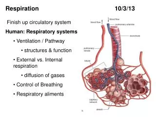
Pulmonaryartery Aorta Pulmonaryartery Superiorvena cava LEFTATRIUM RIGHTATRIUM Pulmonaryveins Pulmonaryveins Semilunarvalve Semilunarvalve Atrioventricularvalve Atrioventricularvalve Inferiorvena cava RIGHTVENTRICLE LEFTVENTRICLE Figure 23.4A
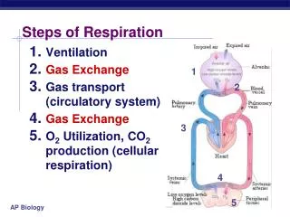
RBC Pathway through the Circulatory System Blood from Systemic Circuit Vena cava (inferior & superior) Right atrium (Tricuspid valve-AV valve) Right ventricle (Pulmonary semilunar valve) Pulmonary circuit –Lungs (P. arteries LungsP. veins) Left atrium (Bicuspid “Mitral” valve) Left Ventricle (Aortic semilunar valve) Aorta (arch, coronary, carotid, & abdominal, renal, mesenteric, iliac arteries)
Introductory Questions #1 • The heart has four valves in it. Name them. Name the blood vessel that carries oxygen poor blood from the heart to the lungs to pick up more oxygen. • Name the two large veins that bring blood to the heart from the rest of the body. • In your textbook (pg. 975) Name the four components of blood. What % of your blood is composed of red blood cells? What about white blood cells?
IQ #2 Pulmonaryartery 11. vessel Aorta 10. vessel Pulmonaryartery 1. vessel Superiorvena cava LEFTATRIUM 2. chamber RIGHTATRIUM Pulmonaryveins Pulmonaryveins 9. vessels 3. valve Semilunarvalve Semilunarvalve 8. valve Atrioventricularvalve 7. valve Atrioventricularvalve 4. vessel Inferiorvena cava 5. chamber RIGHTVENTRICLE 6. chamber LEFTVENTRICLE Figure 23.4A
Observation of a Cow’s Heart • Make a drawing of your section you received-ID it as the anterior or posterior section. • Make labels and pin your specimen • Include these labels on your drawing. • Optional-Take a picture of your labeled specimen. Labels: -Septum -Left & Right Ventricle -Left & Right Atrium -Aorta -AV valve (tricuspid & Bicuspid) -Semilunar valve (pulmonary & aortic)
Video #1: Circulation: River of Life (Ch. 37.2) • What is the primary function of the circulatory system? • How is an open circulatory system different a closed? Give an example of an organism that has an open circulatory system. • How is a vein different from an artery? Give two differences. • Name FOUR chambers and the four valves within the heart. **Write the title for this segment and give FIVE statements.
Pulmonaryartery 11. vessel Aorta 10. vessel Pulmonaryartery 1. vessel Superiorvena cava LEFTATRIUM 2. chamber RIGHTATRIUM Pulmonaryveins 9. vessels Pulmonaryveins 3. valve Semilunarvalve 8. valve Semilunarvalve Atrioventricularvalve 7. valve Atrioventricularvalve 4. vessel Inferiorvena cava 5. chamber RIGHTVENTRICLE 6. chamber LEFTVENTRICLE Figure 23.4A
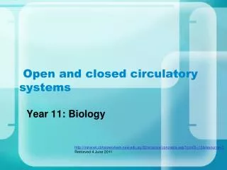
What is a heart attack? • A heart attack is damage that occurs when a coronary feeding the heart is blocked Aorta Rightcoronaryartery Leftcoronaryartery Blockage Dead muscle tissue Figure 23.8A
Reading Assignment • Using the handout read and review some of the key aspects of the circulatory system. • On a separate sheet paper answer Questions on Pgs 25 & 26 from the second handout.
Label & Color your Hear Diagram • Be sure to use RED for all areas that contains oxygen rich blood and BLUE for areas with oxygen poor blood. • All valves must be correctly labeled
Double Circulation • From right ventricle to lungs via pulmonary arteries through semilunar valve (pulmonary circulation) • Capillary beds in lungs to left atrium via pulmonary veins • Left atrium to left ventricle (through atrioventricular valve) to aorta • Aorta to coronary arteries; then systemic circulation • Back to heart via two venae cavae (superior and inferior); right atrium
Introductory Questions #1 • The heart has four valves in it. Name them. Name the blood vessel that carries oxygen poor blood from the heart to the lungs to pick up more oxygen. • Name the two large veins that bring blood to the heart from the rest of the body. • In your textbook (pg. 975) Name the four components of blood. What % of your blood is composed of red blood cells? What about white blood cells?
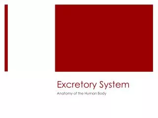
Pg. 880 Withdrawblood Centrifuge Place in tube PLASMA 55% CONSTITUENT MAJOR FUNCTIONS CELLULAR ELEMENTS 45% Solvent forcarrying othersubstances CELL TYPE NUMBER(per mm3 of blood) FUNCTIONS Water Erythrocytes(red blood cells) Salts 5–6 million Transport ofoxygen (and carbon dioxide) Sodium Potassium Calcium Magnesium Chloride Bicarbonate Osmotic balance,pH buffering, andregulation ofmembranepermeability Leukocytes(white blood cells) Defense andimmunity 5,000–10,000 Plasma proteins Albumin Fibrinogen Immunoglobins(antibodies) Osmotic balance,pH buffering Clotting Immunity Lymphocyte Basophil Eosinophil Substances transported by blood Monocyte Nutrients (e.g., glucose, fatty acids, vitamins) Waste products of metabolism Respiratory gases (O2 and CO2) Hormones Neutrophil Platelets 250,000–400,000 Blood clotting Figure 23.13
IQ #2 Pulmonaryartery 11. vessel Aorta 10. vessel Pulmonaryartery 1. vessel Superiorvena cava LEFTATRIUM 2. chamber RIGHTATRIUM Pulmonaryveins Pulmonaryveins 9. vessels 3. valve Semilunarvalve Semilunarvalve 8. valve Atrioventricularvalve 7. valve Atrioventricularvalve 4. vessel Inferiorvena cava 5. chamber RIGHTVENTRICLE 6. chamber LEFTVENTRICLE Figure 23.4A
Introductory Questions #3 • Name the cell fragments that aide in the process of blood clotting. (pg. 977) • What substances are found in the plasma of blood? (pg. 975) • Name the proteins that are found on the surface of red blood cells.
Introductory Questions #4 • Give three differences between arteries and veins. • Why must blood slow down as it reaches a capillary bed? • Where in the heart is the “pacemaker” and what role does it serve?
The Heart Contracts and Relaxes Rhythmically 1 Heart isrelaxed.AV valvesare open. 2 Atriacontract. • Diastole • Blood flows from the veins into the heart chambers • Systole • The atria briefly contract and fill the ventricles with blood • Then the ventricles contract and propel blood out SYSTOLE 0.1 sec 3 Ventriclescontract.Semilunarvalvesare open. 0.3 sec 0.4 sec DIASTOLE Figure 23.6
The Heartbeat • Sinoatrial (SA) node (“pacemaker”): sets rate and timing of cardiac contraction by generating electrical signals • Atrioventricular (AV) node: relay point (0.1 second delay) spreading impulse to walls of ventricles • Electrocardiogram (ECG or EKG)
Velocity of Blood • Aorta: 30cm/sec on average • Capillary: 0.026 cm/sec • Change is caused by the Law of Continuity • As a pipe’s diameter narrows, the flow rate increases • The total cross-sectional area of the capillaries determines flow rate • Each artery branches extensively to an enormous number of capillaries that has a huge cross sectional area that is much greater than the artery, so the flow rate is slowed.
No substance has to diffuse far to enter or leave a cell Capillary Diffusion ofmolecules INTERSTITIALFLUID Tissuecell Figure 23.1B
Pg. 880 Withdrawblood Centrifuge Place in tube PLASMA 55% CONSTITUENT MAJOR FUNCTIONS CELLULAR ELEMENTS 45% Solvent forcarrying othersubstances CELL TYPE NUMBER(per mm3 of blood) FUNCTIONS Water Erythrocytes(red blood cells) Salts 5–6 million Transport ofoxygen (and carbon dioxide) Sodium Potassium Calcium Magnesium Chloride Bicarbonate Osmotic balance,pH buffering, andregulation ofmembranepermeability Leukocytes(white blood cells) Defense andimmunity 5,000–10,000 Plasma proteins Albumin Fibrinogen Immunoglobins(antibodies) Osmotic balance,pH buffering Clotting Immunity Lymphocyte Basophil Eosinophil Substances transported by blood Monocyte Nutrients (e.g., glucose, fatty acids, vitamins) Waste products of metabolism Respiratory gases (O2 and CO2) Hormones Neutrophil Platelets 250,000–400,000 Blood clotting Figure 23.13
SSR Guide for Presenters • Give your name • Hold up the your source • FIVE key points with a brief summary of what you read. • The rest of us need to write points discussed in the SSR boxes on your cover sheet.
Assignment Packet • Cover sheet • Circulatory System Handout Questions • Video Notes (x3) • Heart Diagram (ID &* Color) • Mini-lab Activity: Cow’s Heart (w/stamp)
Introductory Questions #1 • What type of instrument is used to measure a person’s blood pressure? Why is knowing a person’s blood pressure important? • What is “normal” or average blood pressure at rest? What about heart rate? • What does the top number and bottom number represent when reading blood pressure?
Systolicpressure Diastolicpressure • Pressure is highest in the arteries • It drops to zero by the time the blood reaches the veins Relative sizes andnumbersof blood vessels Figure 23.9A
Velocity of Blood • Aorta: 30cm/sec on average • Capillary: 0.026 cm/sec • Change is caused by the Law of Continuity • As a pipe’s diameter narrows, the flow rate increases • The total cross-sectional area of the capillaries determines flow rate • Each artery branches extensively to an enormous number of capillaries that has a huge cross sectional area that is much greater than the artery, so the flow rate is slowed.
Blood Vessel Structural Differences • Capillaries •endothelium; basement membrane • Arteries •thick connective tissue; thick smooth muscle; endothelium; basement membrane • Veins•thin connective tissue; thin smooth muscle; endothelium; basement membrane
Match the Blood Vessels with its location in the Body • Carotid artery Heart • Jugular vein Liver • Coronary Stomach • Hepatic Kidney • Renal Neck (2) • Iliac Arm • Brachial Thigh • Femoral Pelvic • Gastric
Measuring Blood Pressure can Reveal Cardiovascular Problems • Blood pressure is measured as systolic and diastolic pressures Sphygmomanometer Blood pressure120 systolic80 diastolic(to be measured) Pressurein cuffabove120 Pressurein cuffbelow120 Pressurein cuffbelow 80 Rubber cuffinflated with air Soundsaudible instethoscope Soundsstop Arteryclosed Artery 2 3 4 1 Figure 23.10
Lab Activity-Measuring Blood Pressure & Heart Rate Answer to Part A (Fill in Blanks) • Arterial • Systolic • Diastolic • Heart • Blood pressure • Spygmomanometer • Mm Hg • Systolic • Pulse pressure (40mmHg) • Brachial
Today’s Activities • Start IQ #3 ****stamp Hmwk: Pg. 974 #1-4 • Discuss the homework • Discuss Lab Questions from yesterday • Lect/Disc: Respiratory system • Answer IQ #2 & IQ #3 **Begin Video #1: The Respiratory System
Introductory Questions #3 • Name the two types of muscles that allow you to breath in and out. (pg. 973) • When a person inhales, what happens to the diaphragm? What about when you exhale? • When the space in the chest cavity (volume) increases which way does the air move? The textbook states that it creates a slight ____________. (see pg. 974)
Introductory Questions #2 • Reading pg. 971 (Ch. 37), place these terms in the correct order that tracks air as you breath: -Pharynx -nose & mouth -bronchi -alveoli -bronchioles -trachea • How does your body protect itself from the millions of particles in the air as you breath? (see pg. 972) • What are the tiny alveoli sacs surrounded by?
Larynx (upper part of respiratory tract) Vocal cords (sound production) Trachea (windpipe) Bronchi (tube to lungs) Bronchioles Alveoli (air sacs) Diaphragm (breathing muscle) Mammalian Respiratory Systems
The bronchioles end in clusters of tiny sacs called alveoli • Alveoli form the respiratory surface of the lungs • Oxygen diffuses through the thin walls of the alveoli into the blood Figure 22.6C Oxygen-richblood Oxygen-poorblood Bronchiole Alveoli Blood capillaries Figure 22.6B
Breathing • Positive pressure breathing: pushes air into lungs (frog) • Negative pressure breathing: pulls air into lungs (mammals) • Inhalation: diaphragm contraction; Exhalation: diaphragm relaxation • Tidal volume: amount of air inhaled and exhaled with each breath (500ml) • Vital capacity: maximum tidal volume during forced breathing Regulation: CO2 concentration in blood (medulla oblongata)
Smoking causes lung cancer and contributes to heart disease • Smoking also causes emphysema • Cigarette smoke makes alveoli brittle, causing them to rupture • This reduces thelungs’ capacity for gas exchange Figure 22.7A, B
The human respiratory system Nasalcavity Pharynx (Esophagus) Left lung Larynx Trachea Rightlung Bronchus Bronchiole Diaphragm (Heart) Figure 22.6A