Download
1 / 33
390 likes | 922 Views
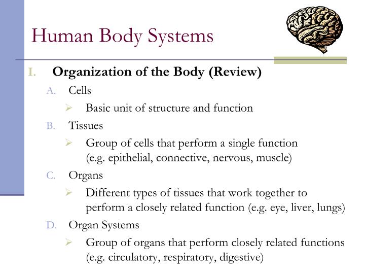
Human Body Systems. Organization of the Body (Review) Cells Basic unit of structure and function Tissues Group of cells that perform a single function (e.g. epithelial, connective, nervous, muscle) Organs

E N D
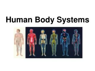
Human Body Systems • Organization of the Body (Review) • Cells • Basic unit of structure and function • Tissues • Group of cells that perform a single function (e.g. epithelial, connective, nervous, muscle) • Organs • Different types of tissues that work together to perform a closely related function (e.g. eye, liver, lungs) • Organ Systems • Group of organs that perform closely related functions (e.g. circulatory, respiratory, digestive)
Homeostasis • Definition: the process by which organisms keep internal conditions relatively constant despite changes in their external environments • Requires the integration of all organ systems at the same time • Nervous system in conjunction with the endocrine system (hormones) is responsible for this integration
Examples of Feedback Inhibition • III. Maintaining Homeostasis • Negative feedback – your body’s response results in decreasing the effect of the stimulus (e.g. body temperature) Section 35-1 Sensed by Room Temperature Drops Room temp. rises Thermostat Signals Heater to turn on
Positive feedback – your body’s response results in an increase in the effect of the stimulus, (e.g. the flight-fight response)
Nervous System • Recognizes and coordinates the body’s response to changes in its internal and external environments. • General Functions of the Nervous System • Sensory input – vision, hearing, balance, smell, taste, and touch • Motor output – muscle contraction and movement • Memory and integration of information
Organization of the NS Central N.S. 1. Brain 2. Spinal Cord B. Peripheral N.S. 1. Somatic N.S. 2. Autonomic N.S. a. Sympathetic b. Parasympathetic
Nervous System • Division of Labor • Central Nervous System (CNS) • Control center of the body that relays messages, and processes and analyzes information • Brain • Cerebrum – largest region; right and left hemispheres that are connected by corpus callosum; voluntary activities and higher brain functions • Cerebellum – located at the lower back part of brain; coordination and balance
Nervous System • Brain stem – connects the brain and spinal chord; two regions: pons and medulla oblongata, control breathing, heart rate and swallowing • Thalamus and hypothalamus - between brain stem and cerebrum Thalamus: relay station for sensory info Hypothalamus: most important homeostatic site; hormones, body’s thermostat, fight or flight, thirst, hunger, reproduction
Draw Fig. 35-9: The Brain Cerebrum Thalamus Pineal gland Hypothalamus Cerebellum Pituitary gland Pons Spinal cord Medulla oblongata
Nervous System • Spinal Cord • Two main fxns: • Processing of simple responses to certain stimuli (reflexes) • Carries info to and from brain to body
Nervous System • Peripheral Nervous System (PNS) • Receives information from the environment and relays to and from CNS and sensory, motor and gland cells
Nervous System • Two divisions: • Sensory - Made of sensory neurons that bring info to the CNS • Motor - Made of sensory neurons that convey info from the CNS; two subdivisions • Somatic (voluntary): respond to external stimuli • Autonomic (involuntary): respond to internal stimuli w/the parasympathetic and sympathetic divisions • Sympathetic ↑energy consumption • Parasympathetic ↓energy consumption
Nervous System • Neurons (Nerve Cells) • Specialized cells that carry electrical signals called impulses (Draw Fig. 35-5; pg. 897) • 3 Types of Neurons: • Sensory – carry impulses from the sense organs to the spinal cord and brain • Motor – carry impulses from brain and spinal cord to muscles and glands • Interneurons – Connect sensory and motor neurons and carry impulses between them
Nervous System • Anatomy of a Neuron • Cell Body • Largest part of the neuron • Contains the nucleus and most of the cytoplasm • Metabolic activity takes place in the cell body Cell Body
Nervous System • Dendrites • Carry impulses from the environment or other neurons to the cell body
Nervous System • Axon • Long fiber that carries impulses from the cell body • Ends in axon terminals that contain vesicles for neurotransmitters
Nervous System • Myelin Sheath • Insulates the axon • Gaps in the myelin sheath allow an impulse to jump from node to node, thus increasing its speed
The Nerve Impulse • The Resting Neuron • At rest, the outside of the cell has a net positive charge and the inside has a netnegative charge. This charge difference is called the resting potential. (-70mVolts, about 5% of the voltage in AA battery)
The Resting Neuron (cont) • The charge difference is created by active transport of ions across the cell membrane via the sodium-potassium pump. • Sodium ions (Na+) are pumped outside the cell and potassium (K+) ions are pumped into the cell.
The Moving Impulse • An impulse begins when a neuron is stimulated by the axon of another neuron or by the environment. • Na+ pores open and the flood of Na+ ions makes the inside positive. _ + + _
The Moving Impulse (cont) • This reversal of charges, from negative to positive is called a nerve impulse, or an action potential. • As the impulse passes, K+ pores open and K+ flows out which restores the resting potential (charge difference) + _ + _
The Moving Impulse (cont) • How do things get back to the original condition? The sodium potassium pump kicks in. • The minimum level of a stimulus that is required to activate a neuron is called the threshold.
Nerve Impulse Pathway Overview • Impulse is received by the dendrites from the environment or another neuron, then gets rapidly channeled through the cellbody to the axon • Axon branches out into axon terminals, which contain tiny vesicles filled with neurotransmitters, which are chemicals used by a neuron to transmit an impulse to another cell. (e.g. acetylcholine, serotonin, dopamine and adrenalin). • Vesicles release neurotransmitters into the open space between neurons called the synapse.
Nerve Impulse Pathway (cont) • The neurotransmitters diffuse across the synapse and attach themselves to receptors on dendrite of neighboring cell Direction of Impulse Dendrite of adjacent neuron Axon Receptor Vesicle Axon terminal Synaptic cleft Neurotransmitter
Nervous System • Reflexes • Reflexes are automatic responses to stimuli • Controlled by 5-part reflex arc: • Sensoryreceptors on finger reacts to stimulus (heat) • Impulse is carried to the spinal cord by a sensoryneuron • In the spinal cord, the impulse is transferred by an interneuron to motor neuron • Motorneurons conducts the impulse to an effector (arm muscles) • Effector responds to the impulses by contracting (hand gets pulled away from the heat)
Nervous System • The Senses 5 General Sensory Receptors: pain, thermo-, mechano-, chemo- and photoreceptors. • Where do you think these different types of receptors are found and what is their function? • Vision • Hearing and Balance • Smell and Taste • Touch
Nervous System • Nervous System Disorders • Migraine Headaches – caused by change in serotonin levels? (affected by caffeine, estrogen, certain foods) • Parkinson’s –caused by damage to dopamine transmitters; causes uncontrollable shaking, no cure • Tay-Sachs –lack enzyme to break down fatty deposits in the brain; neurological deterioration; death by age 4-5 • Dementia - damaged brain cells caused by injury or disease (Alzheimer’s); memory loss and personality change.
Nervous System • Drugs and the Nervous System • Stimulants • Accelerate HR, BP, and breathing rate • Increases the release of neurotransmitters; leads to release of energy and feeling of well-being • When effect wears off, brain’s supply is depleted • Caffeine • Cocaine • Methamphetamines
Nervous System • Depressants • Slow down HR, lower BP and breathing rate, relax muscles and relieves anxiety • Alcohol • Marijuana • Sleeping Pills
Commonly Abused Drugs Commonly Abused Drugs Section 35-5 Used to increase alertness, relieve fatigue Used to relieve anxiety, irritability, tension Used to relieve pain Stimulants Depressants Opiates Amphetamines Barbiturates Tranquilizers Morphine Codeine Increase heart and respiratory rates; elevate blood pressure; dilate pupils; decrease appetite Slow down the actions of the central nervous system; small amounts cause calmness and relaxation; larger amounts cause slurred speech and impaired judgement Act as a depressant; cause drowsiness, restlessness, nausea Drug Type Medical Use Examples Effects on the body