Download
1 / 49
810 likes | 1.47k Views
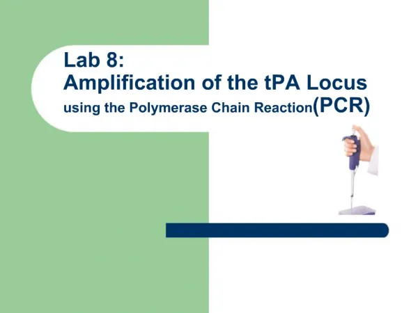
Class 26_2011 updated Dec. 8, 201 12:45 AM. Recombinase Polymerase Amplification. Radhika Pradhan Aidan Quinn Ziwei Song. TwistDx Ltd. qPCR Primer Design. Portable real-time fluorometer. Advantages and Potential Applications. Advantages.

E N D
Class 26_2011 updated Dec. 8, 201 12:45 AM Recombinase Polymerase Amplification RadhikaPradhan Aidan Quinn Ziwei Song TwistDx Ltd.
Advantages • From 1 molecule DNA or 10 molecules RNA to detectable levels (billions or trillions) in 5-10 min • Low cost and simple reagents means practical applications are enormous • Multiplexing allows simultaneous detection of multiple targets
Potential applications • Medical diagnosis • Rapid MRSA detection • Microorganism identification • Agriculture • Portable animal health check • Biodefense • Biohazards (anthrax) • Travel & Public health • SARS • Industrial applications • Food industry • Classrooms • Basement labs • …
Class 26_2011 updated Dec. 8, 201 12:45 AM Therapeutic intervention at the level of pre-mRNA splicing • Interfere with improper splicing caused by splice site creation or activation • E.g., beta-thalassemia (R. Kole) in which a splice site has been created by a mutation in a hemoglobin gene • Use complementary DNA or RNA (antisense) • Natural DNA/RNA rapidly degraded: • Use modified bases, sugars: PNA, morpholino, 2’ OMe, • Normally, DNA-RNA hybrids + endogenous RNase H type activity RNA destruction • Modified antisense DNA circumvents this problem • (don’t want mRNA destroyed here, want to correct its splicing.) PNA = peptide nucleic acids
B. Bias alternative splicing ratios Target the unwanted isoform exon-intron joint. e.g., BCL-2 isoforms, one is pro-apoptotic, one anti-apoptotic. The latter is increased in many cancers Target the anti-apoptotic isoform in cancer cells. e.g., GABA-a-gamma-2 receptor (GABA = gamma amino butyric acid, a neurotransmitter) Long and short forms. Long form associated with mental illness. C. Skip offensive exons e.g., nonsense truncations in dystrophin --->
Splicing as a target for disease therapy Nonsense mutation truncates protein Antisense-induced skipping x Expendable exon (e.g., protein with many repeated domains) Exon must be multiple of 3 in length to maintain reading frame after skipping
RNA modification Deoxy, or also can add 2’ MOE -O-CH2-CH2-O-CH3 MOE = methoxyethyl - Phosphorothioate deoxyoligonucleotides
RNA modification for stabilization ase Morpholino instead of deoxyribose or ribose Modified phosphate ase Still base pairs OK!
Even more extreme and more stable: peptide nucleic acids (PNAs) RNA modification B = a nucleic acid base Amide bonds, No ribose PNA = peptide nucleic acid Attached 1 to 4 lysines here Base pairs even better than natural nucleic acids (higher melting temperatures)
Interfere with improper splicing caused by splice site creation or activation Sazani P, et al. and Kole R. Systemically delivered antisense oligomers upregulate gene expression in mouse tissues Nat Biotechnol. 2002 Dec;20(12):1228-33. EGFP: Enhanced green fluorescent protein = model system Antisense “RNA” injected into tail vein, RNA was modified for stability Mutant globin intron has activated splice sites Actin promoter, universally expressed. Induced exon skipping yields green fluorescence
No antisense: Antisense treatment in cell cultures (ex vivo) from themouse with the mutant EGFP gene Control oligo (C)(50 nt downstream)was ineffective. Max. effect = 40%
C. Skip offensive exons Dystrophin gene 2400 kb, mRNA = 14 kb, 79 exons: a giant gene Protein maintains muscle cell membrane integrity Mutation: Duchenne’s muscular dystrophy Some cases (~half) are due to stop codons (nonsense) in a repetitious exon (spectrin-like repeat, length = a multiple of 3) Deliver antisense to the ends of exon with the nonsense mutation in mdx mice (model for Duchenne’s) to promote the skipping of the nonsense-bearing exon and so avoid truncation of the protein . Use AAV (adeno-associated virus) to deliver the antisense gene Measure: mRNA with skipped exon dystrophin protein muscle histochemistry for dystrophin
Use antisense RNA to target the branch point upstream of the offending exon 23 and the donor splice site downstream of the exon. protein mRNA = 3 X 71 79 BP = branch point; SD = splice donor Branch site (consensus = YNYTRAY) Sequences targeted by antisense
U7 promoter Double target synergistic (loop?) (Kole) compl. to branch compl. to splice donor site Consensus binding site for Sm proteins (to target to pre-mRNA) ITR = inverted terminal repeat, characteristic of AAV
Expression of U7 antisense construct RT-PCR transgenic U7 U7SmOPT-A.S. Endog. U7 (slow onset =conclude slow mRNA turnover) 0 2 4 6 13 weeks Splicing assay (RT-PCR) included Skip exon 23, after 2-4 wks. 0 2 4 6 8 13 weeks normal Dystrophin protein (Western)
dystrophin-associated antigens dystrophin Muscle immuno-histochemistry intriguing Normal Untreated mdx Treated mdx Top, middle ,and bottom
RNAi = RNA interference Short double stranded RNA molecules trigger the degradation of the complementary sequence in the cell, and can inhibit translation of the targeted mRNA Their introduction into a cell can greatly reduce any protein whose mRNA is targeted. Inhibition is usually incomplete in mammalian cells, but can be considerable (>90%) Thus “gene knockdown” as opposed to knock-out siRNA = small inhibitory RNAs shRNA = short hairpin RNAs (both strands can be coded by one DNA) asRNA = antisense RNA miRNA = microRNAs ncRNA = noncoding RNA Alternative technologies: Antisense RNA: block translation or splicing Ribozymes: RNAs that cleave other RNAs, sequence specifically
Introduction of long DS RNA into mammalian cells will trigger the “interferon response: Cessation of protein synthesis via activation of PKR (protein kinase RNA-activated), and phosphorylation of eIF2 Global degradation of mRNA (without any sequence specificity, RNase L activation) Spread to neighboring cells (induction and secretion of interferon) Most small DS RNAs do not trigger this response(<30 bp)
mRNA degradation Inhibits translation of an mRNA
Generation of siRNA in vitro Chemical synthesis, annealing of 22-mers (bypasses dicing by Dicer) T7-mediated in vitro transcription of each complementary strand. Anneal to make long DS RNA and transfer to cells. Let Dicer make siRNA in the cell Also, can use controlled RNase to generatefragments (cheaper) Introduce perfect hairpin RNA into cells, let Dicer make siRNA Introduce imperfect hairpin RNA into cells(based on mRNA sequence) and let Dicer make miRNA
Limitations of siRNA silencing in mammalian cells Transient nature of the response (~3 days) Transfection problems (cell type, refractoriness) Can be cell type specific Non-renewable nature of siRNAs ($$)
Potential determinants of efficient siRNA-directed gene silencing siRNA Incorporation into the RNA-inducing silencing complex (RISC); stability in RISC. Base-pairing with mRNA. Cleavage of mRNA. mRNA Base-pairing with siRNA. The position of the siRNA-binding target region. Secondary and tertiary structures in mRNA. Binding of mRNA-associated proteins. The rate of mRNA translation. The number of polysomes that are associated with translating mRNA. The abundance and half-life of mRNA. The subcellular location of mRNA. Delivery Transfection (lipofection, electroporation, hydrodynamic injection (mouse)) Virus infection (esp. lentivirus (e.g., retrovirus like HIV that can integrate into non-dividing cells)
Some applications: Target oncogene Ras V12 (G12V) – silenced mutant ras without silencing the WT allele. Reduced the oncogenic phenotype (soft agar growth, tumor formation in nude mice) T-lymphocytes infected with anti-CCR5 RNA lower levels of this HIV receptor, and lower levels of infection (5-7X) Target an enzyme in mouse ES cells with a hairpin vector, Isolate a knockdown, make a mouse. Mouse shows same knockdown phenotype in its cells. So can target the whole mammalian organism, Just inject a GFP silencer gene into single cell embryos of a GFP mouse: Can find a chimeric GFP mouse with reduced GFP Progeny carry it in the germ line, Get a complete knockdown mouse, without ES cells (easier)
Delivery in an intact organism Hydrodynamic injection (sudden large volume) of straight siRNA (no vector) into the tail vein of a newborn mouse Get silencing of co-injected luciferase vector in a variety of tissues High throughput siRNA for gene discovery C. elegans, 19,000 genes Make a library 17,000 siRNA genes in plasmids in E. Coli. Feed the clones of E. coli to the worms. Look for phenotypes. 1700 genes examined for phenotypes (as of 2005) (e.g., fat metabolism phenotypes found) Identify the genes affected from sequnce of the siRNA
NATURE 428. 2004. p. 431 tsSV40LTag inactivates p53 at 32o but not at 39o. Infect with Hu shRNA lentivirus shRNA library;select cells that grow at 39o. Knocked down genes = those necessary for p53-induced growth arrest. 32o 39o 39o 39o 39o Control Control p16K.D. p53K.D. p533+p16K,K.D. Identified shRNAs
Systemic RNAi: worms, plants, mammals In plants, get permanent post-transcriptional gene silencing (PTGS, transcriptional level) Worms: effect can last though several generations Amplified by reverse transcriptase Influx/efflux via a specific transmembrane protein (in worms) Raisons d’etre? Infection, many viruses go through a DS RNA phase. Repeat element silencing? (1 million Alus, + others half the human genome) Transcribed in either direction, so could form DS RNA, then RNAi inhibits action of SS ‘mRNA”
Discovery of RNA interference using double-stranded RNA Nature (1998) 391: 806 Discovered RNAi as they tracked down the effective agent in antisense experiments (DS RNA contaminating their SS antisense preparations had all the inhibitory activity) Paper characterized by nice controls and variations: Several genes, whole animal phenotype, protein product (GFP), RNA level (in situs) Phenotype of null mutant is specifically mimicked. Introns and promoter sequences ineffective. DS RNA from a different sequence + SS antisense RNA vs. the target: ineffective DS RNA linked (chimeric molecule) to a single stranded portion vs, the target: ineffective Transport of DS RNA between cells and amplification implied.
In situ hybridizations No probe No RNA injected SS antisense RNA DS RNA Transcript disappears (RNA degraded)
Nucleic acid aptamers Aptamers: molecules that bind other molecules with good affinity and specificity Usually these are proteins . . . . But they can also be RNA or DNA. That is, single stranded RNA or DNA molecules can and will fold up into secondary and tertiary structures depending on their sequence. DNA can be synthesized as very large numbers of different (random sequences) Aptamers can be selected from among these molecules based on their ability to bind an immobilized ligand. The tiny fraction found by chance to be able to bind to your favorite ligand can by amplified by PCR (along with background molecules). Re-iteration of the procedure will enrich for the aptamer until they dominate the population. At this point they can be cloned and sequenced. RNA molecules can be selected by synthesizing them from a randomized DNA population using the T7 promoter appended to each DNA molecule. This enrichment procedure is just the SELEX method described earlier for finding the RNA substrate for RNA binding proteins. In this case it’s the same procedure, looked from the opposite point of view: not what RNA will the protein bind best, but what RNA binds the protein best.
20-mer Random 40 20-mer SELEX Have a random 40-mer synthesized, centered between 2 arbitrary 20-mers (PCR sites) 440 = 1024 Practical limit =1015 = ~ 2 nmoles = ~ 50 ug DNA 1015is a large number.Very large (e.g., 500,000 times as many as all the unique 40-mers in the human genome.) These 1015sequences are known as “sequence space” Each DNA molecule of these 1015(or RNA molecule copied from them) can fold into a particular 3-D structure. We know little as yet about these structures. But we can select the molecules that bind to our target by: AFFINITY CHROMATOGRAPHY Previously discussed SELEX in terms of finding the substrate sequence(s) for an RNA binding protein. Here: select an RNA sequence that can bind any particular target of interest (protein, small molecule).
Who’s binding whom? Protein: thrombin (blood protease) RNA thrombin- binding aptamer
e.g., the soluble form of the immobilized affinity column material SELEX: Systematic Evolution of Ligands by Exponential Enrichment . . . for RNA (or DNA) Essential elements:1) Synthesis of randomized DNA sequences 2) In vitro T7-mediated RNA synthesis from DNA 3) Affinity chromatography 4) RT-PCR DNA (1015) RNA Ligand is immobilized here. Small molecule or large molecule DNA RNA RNA
Some examples of aptamer targets Small molecules Zn+2 ATP adenosine cyclic AMP GDP FMN (and an RNA aptamer is found naturally in E.coli) cocaine dopamine amino acids (arginine) porphyrin biotin organic dyes (cibacron blue, malachite green) neutral disaccharides (cellobiose, and cellulose) oligopeptides aminoglycoside antibiotics (tobramycin) Proteins thrombin HIV tat HIV rev Factor IX (clotting factor) VEGF PDGF ricin large glycoproteins such as CD4 anthrax spores (?)
Tobramycin Electrostatic surface map:red= - blue = + Base flap shuts door
One anti-Rev aptamer: binds peptide in alpha-helical conformation Another anti-Rev aptamer: binds peptide in an extended conformation MS2 protein as beta sheet bound via protruding A.A. side chains Hermann, T. and Patel, D.J.2000. Adaptive recognition by nucleic acid aptamers. Science287: 820-825.
Therapeutic use of an aptamer that binds to and inhibits clotting factor IX Rusconi, C.P., Scardino, E., Layzer, J., Pitoc, G.A., Ortel, T.L., Monroe, D., and Sullenger, B.A. 2002. RNA aptamers as reversible antagonists of coagulation factor IXa. Nature419: 90-94. Reading: Factor IX acts together with Factor VIIIa to cleave Factor X, thus activating it in a step in the blood coagulation cascade leading to a clot. Thus inhibition of Factor IX results in inhibition of clot formation. Desirable during an angioplasty, for example. The usual anti-coagulant used in angioplasty is heparin, which has some toxicity and is difficult to control. Inverted T at 3’ end (3’-3’) slows exonucleolytic degradation ( R-3’O-P-O-3’-R-T )
Anti-Factor IX RNA aptamer isolated by SELEX Kd for Factor IX = 0.6 nM F_IXa + F_VIIIa cleaves F_X 4 nM aptamer inhibits this activity +aptamer-PEG, Clotting time increase +aptamer+PEGylation mutant version -aptamer == 1 Conjugate to polyethyleneglycol to increase bloodstream lifetime PEG = polyethyleneglycol polymer, appended to decrease clearance rate.
An antidote to stop the anti-clotting action if a patient begins to bleed. Would be an improvement over heparin. Just use the complementary strand (partial) as an antidote. The 2 strands find each other in the bloodstream! Antidote 5-2 design = the open squares 16-fold excess Anti-coagulant activity duplexed In human plasma free aptamer Scrambled antidote +Oligomer 5-2 Ratio of anti- to aptamer
Antidote acts fast (10 min) Anti-coagulant activity Need 10X antidote Antithrombin aptamer antidote tested in human serum Ratio antidote/aptamer Anti-coagulant activity Time (min) Anti-coagulant activity Antidote lasts a long time Time (hr)
Reduced clotting Reversed by antidote In serum of patients with heparin-induced thrombocytopenia (heparin can no longer be used)
Macugen: an RNA aptamer that binds VEGF and is marketed for adult macular degeneration (wet type) From the label: R Inverted ribo-T 3’-3’ to protect 3’ end Where R is and contains a PEG chain of ~ 450 ethylene glycol units. The chemical name for pegaptanib sodium is as follows: RNA, ((2'-deoxy-2'-fluoro)C-Gm-Gm-A-A-(2'-deoxy-2'-fluoro)U-(2'-deoxy-2'-fluoro)C-Am-Gm-(2'-deoxy-2′-fluoro)U-Gm-Am-Am-(2'-deoxy-2'-fluoro)U-Gm-(2'-deoxy-2'-fluoro)C-(2'-deoxy-2'-fluoro)U-(2'-deoxy-2'-fluoro)U-Am-(2'-deoxy-2'-fluoro)U-Am-(2'-deoxy-2'-fluoro)C-Am-(2'-deoxy-2'-fluoro)U-(2'-deoxy-2'-fluoro)C-(2'-deoxy-2'-fluoro)C-Gm-(3'→3')-dT), 5'-ester with α,α'-[4,12-dioxo-6-[[[5-(phosphoonoxy)pentyl]amino]carbonyl]-3,13-dioxa-5,11-diaza-1,15-pentadecanediyl]bis[ω- methoxypoly(oxy-1,2-ethanediyl)], sodium salt. The molecular formula for pegaptanib sodium is C294H342F13N107Na28O188P28[C2H4O]n (where n is approximately 900) and the molecular weight is approximately 50 kilodaltons. Macugen is formulated to have an osmolality of 280-360 mOsm/Kg, and a pH of 6–7. VEGF = vascular endothelial growth factor