Download
1 / 7
70 likes | 162 Views
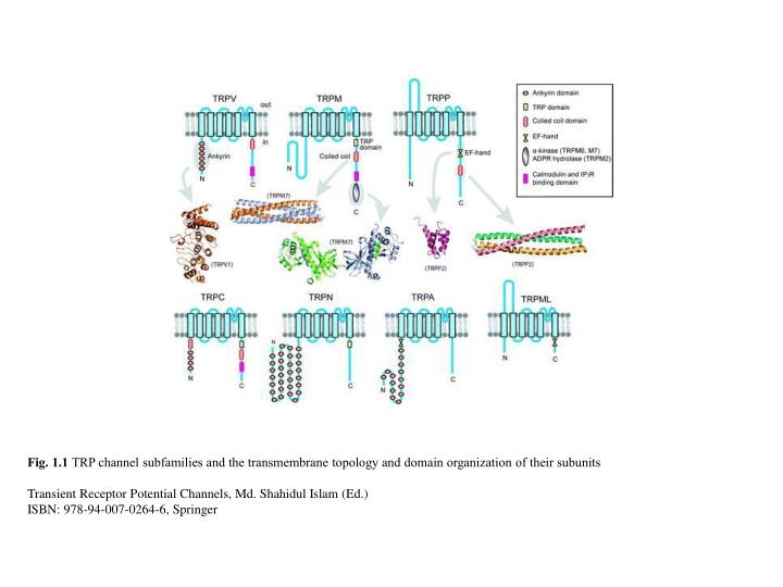
Fig. 1.1 TRP channel subfamilies and the transmembrane topology and domain organization of their subunits Transient Receptor Potential Channels, Md. Shahidul Islam (Ed.) ISBN: 978-94-007-0264-6, Springer.

E N D
Fig. 1.1 TRP channel subfamilies and the transmembrane topology and domain organization of their subunits Transient Receptor Potential Channels, Md. Shahidul Islam (Ed.) ISBN: 978-94-007-0264-6, Springer
Fig. 1.2 Examples of membrane protein structures at different resolutions. (a) Side view (left) and top view (right) of a cryo-EM structure of the Drosophila Shaker K+ channel. (b) Side view of the structure of a monomer of aquaporin 1. (c) X-ray crystal structure of the rat Kv1.2 channel. (d) The electron density map and side chain assignment of the ion selectivity filter of a rat Kv1.2–Kv2.1 chimeric channel Transient Receptor Potential Channels, Md. Shahidul Islam (Ed.) ISBN: 978-94-007-0264-6, Springer
Fig. 1.3 TRP channel EM structures. (a) Cryo-EM structure of TRPV1 [21]. (b) Cryo-EM structure of TRPV4. (c) EM structure of TRPM2 with negative staining [19]. (d) Cryo-EM structure of TRPC3 [20] Transient Receptor Potential Channels, Md. Shahidul Islam (Ed.) ISBN: 978-94-007-0264-6, Springer
Fig. 1.4 Crystal structure of the TRPM7 α-kinase domain. (a) The dimeric structure of the TRPM7 α-kinase domain (PDB code 1IA9). (b) and (c) Comparison of the TRPM7 α-kinase domain (b, PDB code 1IA9) and PKA kinase domain (c, PDB code 1CDK) Transient Receptor Potential Channels, Md. Shahidul Islam (Ed.) ISBN: 978-94-007-0264-6, Springer
Fig. 1.5 Crystal structures of TRPV ankyrin repeat domains (ARDs). (a) Superposition of the structures of the ARD of TRPV1 (PDB code 2PNN), TRPV2 (PDB code 2ETB), TRPV4 (PDB code 3JXI), and TRPV6 (PDB code 2RFA). (b) Structure of the TRPV1-ARD with a bound ATP molecule. (c) Structure of the TRPV4-ARD Transient Receptor Potential Channels, Md. Shahidul Islam (Ed.) ISBN: 978-94-007-0264-6, Springer
Fig. 1.6 Comparison of the crystal structure of a C-terminal coiled coil domain of TRPM7 (a, PDB code 3E7K) and TRPP2 (b, PDB code 3HRN) Transient Receptor Potential Channels, Md. Shahidul Islam (Ed.) ISBN: 978-94-007-0264-6, Springer
Fig. 1.7 NMR structure of an E-F hand in the TRPP2 C terminus Transient Receptor Potential Channels, Md. Shahidul Islam (Ed.) ISBN: 978-94-007-0264-6, Springer