Download

1 / 49
490 likes | 756 Views
The Non-Leukaemic Lymphoproliferative Disorders. Ahmad Sh. Silmi Msc Hematology, FIBMS IUG. The Non-Leukaemic Lymphoproliferative Disorders. These are malignant clonal disorders of the lymphopoietic system and include: Multiple Myeloma and Related Plasma Cell Disorders HD NHL.

E N D
The Non-Leukaemic Lymphoproliferative Disorders Ahmad Sh. Silmi Msc Hematology, FIBMS IUG
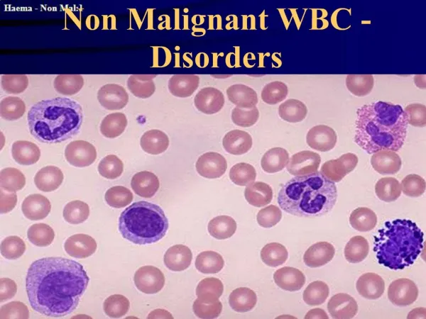
The Non-Leukaemic Lymphoproliferative Disorders These are malignant clonal disorders of the lymphopoietic system and include: • Multiple Myeloma and Related Plasma Cell Disorders • HD • NHL
Multiple Myeloma and Related Plasma Cell Disorders • Multiple Myeloma (MM) is a B lymphoid malignancy which, is characterized by the proliferation of a malignant clone of plasma cells which synthesize and secrete excessive amounts of monoclonal immunoglobulin. In many respects, MM behaves as a solid tumour with a prior involvement for bone marrow.
Incidence • Disease of elderly. • The median age at diagnosis is about 62 years. • The disease is more common in blacks than in whites. • The disease shows slight excess incidence in male than female.
Clinical symptoms • bone pains, pathologic fractures • weakness and fatigue • serious infection • renal failure • bleeding diathesis
Laboratory tests • ESR > 100 • anaemia, thrombocytopenia • rouleaux in peripheral blood smears • marrow plasmacytosis > 10 -15% • hyperproteinemia • hypercalcemia • proteinuria • azotemia
Diagnostic Criteria for Multiple Myeloma Major criteria I. Plasmacytoma on tissue biopsy II. Bone marrow plasma cell > 30% III. Monoclonal M spike on electrophoresis IgG > 3,5g/dl, IgA > 2g/dl, light chain > 1g/dl in 24h urine sample Minor criteria a. Bone marrow plasma cells 10-30% b. M spike but less than above c. Lytic bone lesions d. Normal IgM < 50mg, IgA < 100mg, IgG < 600mg/dl
Diagnostic Criteria for Multiple Myeloma Diagnosis: • I + b, I + c, I + d • II + b, II + c, II + d • III + a, III + c, I II + d • a + b + c, a +b + d
Staging of Multiple Myeloma Clinical staging • is based on level of haemoglobin, serum calcium, immunoglobulins and presence or not of lytic bone lesions • correlates with myeloma burden and prognosis I. Low tumor mass II. Intermediate tumor mass III. High tumor mass • subclassification A - creatinine < 2mg/dl B - creatinine > 2mg/dl
Poor prognosis factors • cytogenetical abnormalities of 11 and 13 chromosomes • beta-2 microglobulines > 2,5 ug/ml
Treatment At present, there is no curative therapy, but symptomatic treatment may be as: • Blood transfusion if anaemia present. • Antibiotic to treat infection. • Local radiotherapy for osteolytic bone. • Dialysis in case of renal failure. • Alkalating agents may provide pain relief, but the response to these drugs is slow.
Waldenstrom's Macroglobulinaemia • Waldenstrom's Macroglobulinaemia (WM) is an uncommon B lymphoid disorder, which is characterized by hyperviscosity secondary to the excessive secretion of a monoclonal IgM immunoglobulin by the malignant clone.
Causes • It is caused by the loss of regulation of a clone of cells, which appear to be in an intermediate stage of development between the mature lymphocytes and the early plasma cells. Morphologically, the malignant cells of WM are rather more immature than those in MM and frequently are described as being "lymphoplasmacytoid".
Incidence • WM is a disease of elderly, with a peak incidence occurring in the seventh decade of life with no sex predilection. • Life expectancy ranges from 8 months to 8 years
Symptoms • Weight loss. • Hepatosplenomegaly. • Lymphadenopathy. • bruising or bleeding tendency. • A long history of vague weakness, fatigue and weight loss. • Hyperviscosity syndrome due to malignant infiltration or accumulation of monoclonal immunoglobulin.
Clinical symptoms Accumulation of the IgM can lead to a variety of clinical symptoms include: • Neurological symptoms such as headache, vertigo, and in severe cases coma. • Visual disturbances secondary to retinal haemorrhage and oedema, which may cause permanent blindness. • Cardiac failure which is severe by the increased plasma viscosity. • Platelet dysfunction secondary to coating of the platelets by the monoclonal IgM. • Haemostatic disturbances secondary to the inhibition of fibrin polymerization and factor VIII activity by the monoclonal IgM.
Laboratory Findings • Normocytic normochromic anaemia secondary to suppression of erythropoiesis by the malignant clone. • Rouleaux formation secondary to hyperviscosity. • Chronic bleeding and dilution by the increased plasma volume. • The WBC may be normal or depressed but a relative lymphocytosis commonly is present. • The platelet count is normal at presentation. However, neutropenia and thrombocytopenia become more severe as the disease progress. • Hypercellular and extremely hyperviscous bone marrow, which makes attempts to aspirate the bone marrow frequently difficult. • The malignant cells are pleomorphic: some resembles lymphocytes whereas others clearly resemble plasma cells; most, however, have an intermediate appearance and are described as being lymphoplasmacytoid.
Prognosis Depends on the pattern of infiltration of the malignant clone in the bone marrow: • Diffuse infiltrationis associated with poor prognosis, with a median survival of 17 months. • Nodular infiltration is associated with a much better prognosis, with a median survival of 72 months. • The intermediate form of infiltration, which shows features of both nodular and diffuse infiltration, is associated with a median survival of 52 months.
Treatment • Symptomatic relief from hyperviscosity syndrome is achieved mostly by repeated plasmapheresis. Progressive disease is treated with chemotherapy.
Hodgkin's Disease • Hodgkin's disease (HD) is aneoplastic disorder with development of specific infiltrate containing pathologic Reed-Sternberg cells. It usually arises in lymph nodes and spreads to contiguous groups. Extranodal presentation are rare. Disease is associated with defective cellular immunity.
Incidence • 2-4 cases per 100000 population / year • bimodal age distribution : 15-35 years and above 50 years • male predominance M:F = 1,7:1
Pathophysiology • The most common presenting feature in HD is the presence of painless, a symmetrical enlargement of cervical or supraclavicular lymph nodes. Axillary, inguinal or femoral lymphadenopathy also is seen occasionally. • The lymphadenopathy may be accompanied by severe, generalized itching in the absence of skin rash. • In contrast to non-Hodgkin's lymphomas, which frequently are disseminated at presentation, most cases of HD are restricted to a single anatomical site at presentation. • The presence of pyrexia and night sweats usually are associated with more advanced disease.
Clinical Presentation • Nontender lymph nodes enlargement ( localised ) • neck and supraclavicular area 60-80% • mediastinal adenopathy 50% • other ( abdominal, extranodal disease ) • systemic symptoms (B symptoms) 30% • fever • night sweats • unexplained weight loss (10% per 6 months) • other symptoms • fatigue, weakness, pruritus • cough , chest pain, shortness of breath, vena cava syndrome • abdominal pain, bowel disturbances, ascites • bone pain
Recognition • The peripheral blood is entirely normal at presentation. • Occasionally, mild non-specific changes such as; a mild thrombocytosis, neutropenia or relative eosinophilia is present. • The presence of anaemia, lymphocytopenia or leucoerythroblastosis all suggest the presence of advanced disease with bone marrow involvement, but this is uncommon at presentation. • The disease is recognized by histological examination of an affected lymph node biopsy, which reveals the presence of a diffuse infiltrate of lymphocytes, histiocytes, and eosinophil, plasma cells and neutrophils, which are of normal appearance. Scattered among this infiltrate are variable numbers of Reed-Sternberg (RS) cells, the characteristic feature of HD. • The presence of RS cells is not specific for HD, they also are present in some cases of infectious mononucleosis, NHL, and CLL but their demonstration is required for a diagnosis of HD. • RS cells typically are large, with two or more large, oval nuclei, each of which contains a huge nucleolus, which is separated from the thickened nuclear membrane by a clear zone.
Classification On the basis of the pattern of the lymph node infiltration, four subtypes of HD are recognized:
Lymphocyte predominant HD (LPHD) is characterized by: • A heavy infiltrate of small lymphocytes and histiocytes which have a normal morphology. • The infiltrate is diffuse but may form loose nodules. • RS cells usually are sparse. • This subtype of HD is common in young men. • It's associated with a rapid response to treatment and good prognosis.
Nodular sclerosing HD (NSHD) Involves: • Nodular sclerosis and the presence of classical RS cells, as well as a distinctive RS cell variant called Lacunar cell in which the cell cytoplasm has contracted as an artifact of fixation, leaving an unstained zone between it and the surrounding tissue. • Sclerosis is found in the form of well-organized bands of collagen that subdivides the tissue into distinct nodules. • NSHD is the most common subtype, accounting for more than 40% of cases. • It appears to be commonly associated with a thymic origin and offers a fairly good prognosis.
Mixed cellularity HD (MCHD) is characterized by: • The presence of large numbers of typical and mononuclear RS cells, scattered among morphologically normal lymphocytes, histiocytes, neutrophils, eosinophils, plasma cells and fibroblasts. • This subtype of HD is associated with a less favorable prognosis than either of the above subtypes.
Lymphocyte depleted HD (LDHD) • Large numbers of RS cells and atypical histiocytes, scanty lymphocytes and variable fibrosis. • This subtype is the least common form of HD, and is associated with elderly subjects, who often present with advanced disease and have a poor prognosis.
Immunophenotyping The consistent antigenic markers on RS cells include: • CD25, the IL-2 receptor, CD15 and CD30. • The NSHD and MCHD are more commonly associated with B lymphoid markers while LPHD is associated with T lymphoid markers.
Treatment • According to the stage of disease, early stage requires localized radiotherapy to the involved lymph nodes, while advanced stage involve the use of combination chemotherapy with or without radiotherapy.
Prognosis • With modern treatment, more than 80% of cases of stage I HD and more than 50% of stage IV HD survive for more than 10 years after presentation. Most of these can be considered to be cured.
Aetiology • Possible infectious aetiology secondary to EBV. This virus induces chronic suppression of the immune system, such as occurs in AIDS. Recent evidence has suggested that reactivation of EBV is associated with an increase incidence of NHL. • Epidemiological studies have shown a small excess of NHL among agricultural workers and rubber industry workers but the significance of this observation remains in doubt. • Cytogenetic abnormalities are present in almost all cases of NHL. The most common chromosomal rearrangement often involves chromosome2, 3, 7, 12, 14 and 18.
Pathophysiology • Lymphomas with a follicular pattern of growth are less aggressive than those with a diffuse pattern of growth. • Small lymphocytic lymphomas are less aggressive than large cell lymphomas. • In common with acute leukaemias, some forms of high-grade lymphoma are more amenable to treatment than the chronic types. • In contrast to HD, most NHL is disseminated to a greater or lesser extent at presentation.
Classification • Low Grade Lymphoma • Intermediate Grade Lymphoma • High Grade Lymphoma
Treatment • Lymphomas with a follicular pattern of growth are less aggressive than those with a diffuse pattern of growth. • Small lymphocytic lymphomas are less aggressive than large cell lymphomas. • In common with acute leukaemias, some forms of high-grade lymphoma are more amenable to treatment than the chronic types. • In contrast to HD, most NHL is disseminated to a greater or lesser extent at presentation.