Download
1 / 16
180 likes | 1.13k Views
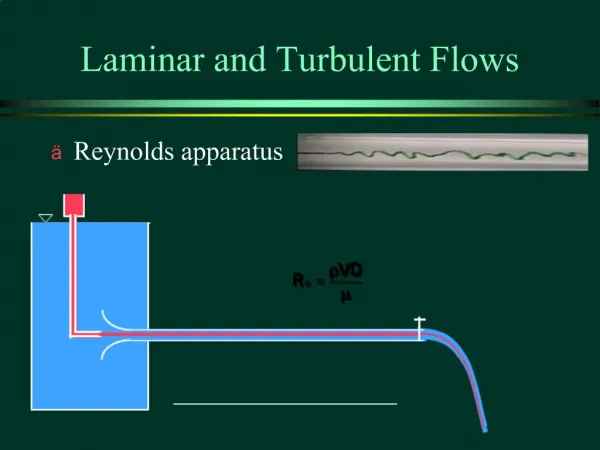
Turbulent Arterial Flows. Mike Fortin. Definitions and Nomenclature. Pulsatile Flow Stenosis Throat Systole Diastole. Scale. Diameter of an Artery Aorta ~ 25 mm Capillary ~ 8 μm Velocity in an Artery Aorta ~ 0.3-0.5 m/s Capillary ~ 0.00-0.01 m/s Reynolds Numbers 1-4000.

E N D
Turbulent Arterial Flows Mike Fortin
Definitions and Nomenclature • Pulsatile Flow • Stenosis • Throat • Systole • Diastole
Scale • Diameter of an Artery • Aorta ~ 25 mm • Capillary ~ 8 μm • Velocity in an Artery • Aorta ~ 0.3-0.5 m/s • Capillary ~ 0.00-0.01 m/s • Reynolds Numbers 1-4000
Specific Arteries • Abdominal Aortic • Reynolds Number ~ 600 in a normal healthy individual. • Can increase to ~ 6000 in the same individual during exercise conditions.
What is in Blood? • Proteins, lipoproteins, ions, and cells. • Red blood cells • Red blood cells makeup approximately 40% of blood by volume. • Semisolid particles. • Increase blood viscosity • Cause non-Newtonian behavior in small blood vessels.
Turbulence Modeling • What model works the best? • Many have been tested. • Large Eddy Simulation? • Time-averaged Navier-Stokes? • Computationally intensive. Not Practical.
Problems with Modeling • Blood is a non-Newtonian fluid in small vessels. In larger vessels, this can be neglected. • Arteries are somewhat elastic. Diameter is not constant. • Arteries dilate to accommodate for increases in flow. • Contraction occurs to control systemic vascular resistance or venous pooling. • Many physical attempts at modeling use rigid tubes.
Model Validation • X-ray contrast angiography • Requires injection of radioactive substance • Tells percent stenosis, but nothing of flow rate • Doppler ultrasound • Require an acoustic or optical window • Tell velocities within 90% • Magnetic resonance imaging • Turbulence in a stenosis causes signal loss
Arteries, but not Veins? • Pulsatile flow exists in arteries. Veins exhibit a fairly constant flow. • Blood flow in arteries is coming from the heart. • Blood flow in veins is going to the heart. • By the time blood gets to the veins, losses have created a fairly steady flow.
Turbulence at a Bifurcation • Carotid artery bifurcation • Flow is mostly laminar entering the division • Separation occurs and turbulence develops * American Heart Association
Stenosis • Flow separation occurs at low Reynolds Numbers • With a 70% stenosis, critical upstream Reynolds Number is only 300. * Churchill Livingstone INC
Stenosis Effects on Flow • Diameter of the vessel decreases • Typically given in a percent reduction in diameter. • Velocity increase is not linear with percent decrease of diameter. It increases as the square. • Causes flow separation. • Turbulence downstream causes significant flow resistance.
Is This Bad? • “Most flows are turbulent. Laminar flow is the exception.” • The body likes laminar flow. • Turbulent flow reduces pressure and causes head loss.
How Bad is Bad? • Aneurysms • Abdominal Aortic ~ 15,000 deaths per year in the US. • Stroke • Heart Attack • Lower-Limb Ischemia • Replacement of Blood Vessels • Must be the same size
Conclusions • Medical community • Not engineers. Not necessarily content, but have sufficient knowledge to know turbulence is not a good thing. • Engineering community • One more area that needs to be advanced to the point where a model is reliable enough to predict the laminar-turbulent transition and the turbulent blood flow regime.
References • Ku, David N. “Blood Flow in Arteries.” Annual Review of Fluid Mechanics. 1997. 29:399-434. • Taylor, C.A., Hughes, T.J.R., Zarins, C.K. “Finite Element Modeling of Three Dimensional Pulsatile Flow in the Abdominal Aorta: Relevance to Atherosclerosis. Annals of Biomedical Engineering, 1998. 26:975-987. • Yellin, E.L., “Laminar-Turbulent Transition Process in Pulsatile Flow.” Circulation Research, 1966. XIX:791-804. • Younis, B.A., Berger, S.A. “ A Turbulence Model for Pulsatile Arterial Flows.” ASME. 2004. 126:578-584.