Download
1 / 16
220 likes | 759 Views
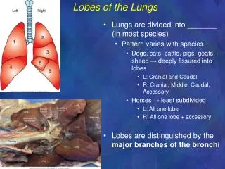
Assessment of the Thorax and Lungs. NUR123 Spring 2009 K. Burger, MSEd, MSN, RN, CNE PPP by: Victoria Siegel RN, MSN, CNS Sharon Niggemeier RN, MSN Revised by: Kathleen Burger. Anatomy of Lungs. Organs of respiration Located in thoracic cavity Right lung-3 lobes Left lung- 2 lobes

E N D
Assessment of the Thorax and Lungs NUR123 Spring 2009 K. Burger, MSEd, MSN, RN, CNE PPP by: Victoria Siegel RN, MSN, CNS Sharon Niggemeier RN, MSN Revised by: Kathleen Burger
Anatomy of Lungs • Organs of respiration • Located in thoracic cavity • Right lung-3 lobes • Left lung- 2 lobes • Important to know landmarks of thorax • Composed of trachea, bronchioles & alveoli
Anatomical Landmarks • Anteriorly: Apex of lung ¾ -1 and ½” (2-4cm) above clavicle. • Anteriorly: Base to 6th rib midclavicular, 8th rib midaxillary. • Posterior: Apex- first thoracic vertebrae. • Posterior: Base T-10 expiration and T -12 deep inspiration.
Subjective Data • Cough • Sputum • Shortness of Breath (SOB) • Smoking • Past Hx respiratory disorders • Environmental factors
Inspection of Thorax and Lungs • With patient sitting up- uncovered • Observe for lesions, chest symmetry, ventilatory pattern, depth, rate and rhythm, muscles used & skin color • Note both posterior view and anterior view. • Note spinal deformities
Palpation of Posterior Thorax • Using fingers palpate chest wall note: • Tenderness • Alignment • Any Bulging or retractions • Palpate for masses • Palpate for any crepitus- coarse, crackling sensation palpable over skin surface in subcutaneous emphysema. May follow thoracic injury or surgery.
Palpate Tactile Fremitus • First say “ahhhh” and feel own neck = fremitus. • Palpate the patient’s back to right and left of spine as the pt. says 99 and examiner palpates with palm of hand, compare bilaterally.
Palpate Chest Expansion/Excursion • Posterior- place hands along outer edge of costal margin with thumbs toward middle of spine • Have patient take a deep breath • Should observe yours hands moving equally far apart.
Percuss the Thorax • Apices to bases • Anterior • Lateral • Posterior- fold arms across chest • Hear resonance and dullness alternately with lung or ribs. • Avoid percussion over scapulae and ribs.
Diaphragmatic Excursion • Distance between deep inspiration and full expiration. • Normally ranges from 3-6 cm • Exhale and hold, percuss and mark location of diaphragm: change dull-resonance • Deep inspiration and hold it, percuss + mark change again
Auscultation • Beginning at apices to base, compare bilaterally. • Listen for full cycle, note quality and intensity • Instruct patient to breathe through mouth, a little deeper (but not faster) than usual • Use stethoscope diaphragm firmly vs chest wall
Normal Breath Sounds • Bronchial- heard over trachea and larynx. High pitch, loud, harsh. Inspiration < expiration • Bronchovesicular- heard over major bronchi. Moderate pitch and loudness.Inspiration=expiration • Vesicular- heard over lung fields. Low pitch, soft sound. Inspiration>expiration
Adventitious sounds • Crackles- (rales) rub hair between fingers cracking/popping sound. Secondary to fluid in airway or to opening of collapsed alveoli in atelectasis. • Wheezes- continuous musical and high pitched, due to constricted bronchi. • Rhonchi- lower pitched, coarse, snoring, due to thick secretions. • Pleural friction rub- rough, grating, inflamed surfaces, as in pleurisy.
Assess Lungs • Note:decreased or absent breath sounds • Bronchial tree obstructed at some point by secretions, mucus plug or foreign body • Emphysema • Anything that obstructs sound transmission: pleurisy, pleural thickening, air (pneumothorax), fluid (pleural effusion), in pleural space.
Increased Breath Sounds • Sounds are louder than they should be, e.g., bronchial sounds heard over peripheral lung fields. • They occur when consolidation e.g., pneumonia or compression creates a denser lung area that enhances sound transmission.
Further Assessment • Bronchophony- say “99”, if heard loud and distinct, it is abnormal • Whispered pectoriloquy- whisper “1,2,3”should be muffled. Abnormal= loud &distinct means there is consolidation. • Egophony – say “E”, the E changes to an “A” sound over area of consolidation, pleural effusion or abscess.