Download
1 / 1
10 likes | 151 Views
ISOLATION AND IDENTIFICATION OF SELENITE REDUCING ARCHAEA FROM TUZ (SALT) LAKE IN TURKEY GUVEN, K., M. B. MUTLU, and C. ÇIRPAN Anadolu University, Faculty of Science Department of Biology Eskisehir, Turkey Email:kguven@anadolu.edu.tr. INTRODUCTION

E N D
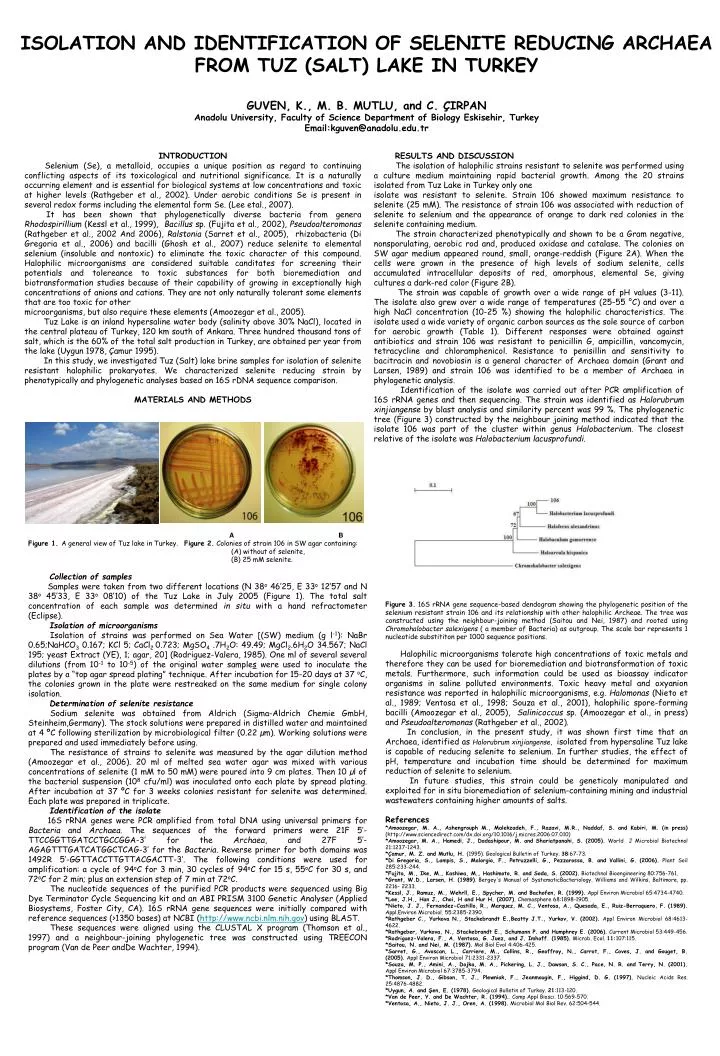
ISOLATION AND IDENTIFICATION OF SELENITE REDUCING ARCHAEA FROM TUZ (SALT) LAKE IN TURKEYGUVEN, K., M. B. MUTLU, and C. ÇIRPANAnadolu University, Faculty of Science Department of Biology Eskisehir, TurkeyEmail:kguven@anadolu.edu.tr INTRODUCTION Selenium (Se), a metalloid, occupies a unique position as regard to continuing conflicting aspects of its toxicological and nutritional significance. It is a naturally occurring elementand is essential for biological systems at low concentrations andtoxic at higher levels (Rathgeber et al., 2002). Under aerobic conditions Se is present in several redox forms including the elemental form Se. (Lee etal., 2007). It has been shown that phylogenetically diverse bacteria from genera Rhodospirillium (Kessl et al., 1999), Bacillus sp. (Fujita et al., 2002), Pseudoalteromonas (Rathgeber et al., 2002 And 2006), Ralstonia (Sarret et al., 2005), rhizobacteria (Di Gregoria et al., 2006) and bacilli (Ghosh et al., 2007) reduce selenite to elemental selenium (insoluble and nontoxic) to eliminate the toxic character of this compound. Halophilic microorganisms are considered suitable canditates for screening their potentials and tolereance to toxic substances for both bioremediation and biotransformation studies because of their capability of growing in exceptionally high concentrations of anions and cations. They are not only naturally tolerant some elements that are too toxic for other microorganisms, but also require these elements (Amoozegar et al., 2005). Tuz Lake is an inland hypersaline water body (salinity above 30% NaCl), locatedinthe central plateau of Turkey, 120 km south of Ankara. Three hundred thousand tons of salt, which is the 60% of the total salt production in Turkey, are obtained per year from the lake (Uygun 1978, Çamur 1995). In this study, we investigated Tuz (Salt) lake brine samples for isolation of selenite resistant halophilic prokaryotes. We characterized selenite reducing strain by phenotypically and phylogenetic analyses based on 16S rDNA sequence comparison. MATERIALS AND METHODS RESULTS AND DISCUSSION The isolation of halophilic strains resistant to selenite was performed using a culture medium maintaining rapid bacterial growth. Among the 20 strains isolated from Tuz Lake in Turkey only one isolate was resistant to selenite. Strain 106 showed maximum resistance to selenite (25 mM). The resistance of strain 106 was associated with reduction of selenite to selenium and the appearance of orange to dark red colonies in the selenite containing medium. The strain characterized phenotypically and shown to be a Gram negative, nonsporulating, aerobic rod and, produced oxidase and catalase. The colonies on SW agar medium appeared round, small, orange-reddish (Figure 2A). When the cells were grown in the presence of high levels of sodium selenite, cells accumulated intracellular deposits of red, amorphous, elemental Se, giving cultures a dark-red color (Figure 2B). The strain was capable of growth over a wide range of pH values (3-11). The isolate also grew over a wide range of temperatures (25-55 °C) and over a high NaCl concentration (10-25 %) showing the halophilic characteristics. The isolate used a wide variety of organic carbon sources as the sole source of carbon for aerobic growth (Table 1). Different responses were obtained against antibiotics and strain 106 was resistant to penicillin G, ampicillin, vancomycin, tetracycline and chloramphenicol. Resistance to penisillin and sensitivity to bacitracin and novobiosin is a general character of Archaea domain (Grantand Larsen, 1989) and strain 106 was identified to be a member of Archaea in phylogenetic analysis. Identification of the isolate was carried out after PCR amplification of 16S rRNA genes and then sequencing. The strain was identified as Halorubrum xinjiangense by blast analysis and similarity percent was 99 %. The phylogenetic tree (Figure 3) constructed by the neighbour joining method indicated that the isolate 106 was part of the cluster within genus Halobacterium. The closest relative of the isolate was Halobacterium lacusprofundi. A B Figure 1. A general view of Tuz lake in Turkey. Figure 2. Colonies of strain 106 in SW agar containing: (A) without of selenite, (B) 25 mM selenite. Collection of samples Samples were taken from two different locations (N 38o 46’25, E 33o 12’57 and N 38o 45’33, E 33o 08’10) of the Tuz Lake in July 2005 (Figure 1). The total salt concentration of each sample was determined in situ with a hand refractometer (Eclipse). Isolation of microorganisms Isolation of strains was performed on Sea Water [(SW) medium (g l-1): NaBr 0.65;NaHCO3 0.167; KCl 5; CaCl2 0.723; MgSO4 .7H2O: 49.49; MgCl2.6H2O 34.567; NaCl 195; yeast Extract (YE), 1; agar, 20] (Rodriguez-Valera, 1985). One ml of several several dilutions (from 10-1 to 10-5) of the original water samples were used to inoculate the plates by a “top agar spread plating” technique. After incubation for 15-20 days at 37 oC, the colonies grown in the plate were restreaked on the same medium for single colony isolation. Determination of selenite resistance Sodium selenite was obtained from Aldrich (Sigma-Aldrich Chemie GmbH, Steinheim,Germany). The stock solutions were prepared in distilled water and maintained at 4 ºC following sterilization by microbiological filter (0.22 µm). Working solutions were prepared and used immediately before using. The resistance of strains to selenite was measured by the agar dilution method (Amoozegar et al., 2006). 20 ml of melted sea water agar was mixed with various concentrations of selenite (1 mM to 50 mM) were poured into 9 cm plates. Then 10 µl of the bacterial suspension (108 cfu/ml) was inoculated onto each plate by spread plating. After incubation at 37 ºC for 3 weeks colonies resistant for selenite was determined. Each plate was prepared in triplicate. Identification of the isolate 16S rRNA genes were PCR amplified from total DNA using universal primers for Bacteria and Archaea. The sequences of the forward primers were 21F 5’-TTCCGGTTGATCCTGCCGGA-3’ for the Archaea, and 27F 5’-AGAGTTTGATCATGGCTCAG-3’ for the Bacteria. Reverse primer for both domains was 1492R 5’-GGTTACCTTGTTACGACTT-3’. The following conditions were used for amplification: a cycle of 94oC for 3 min, 30 cycles of 94oC for 15 s, 55oC for 30 s, and 72oC for 2 min; plus an extension step of 7 min at 72oC. The nucleotide sequences of the purified PCR products were sequenced using Big Dye Terminator Cycle Sequencing kit and an ABI PRISM 3100 Genetic Analyser (Applied Biosystems, Foster City, CA). 16S rRNA gene sequences were initially compared with reference sequences (>1350 bases) at NCBI (http://www.ncbi.nlm.nih.gov) using BLAST. These sequences were aligned using the CLUSTAL X program (Thomson et al., 1997) and a neighbour-joining phylogenetic tree was constructed using TREECON program (Van de Peer andDe Wachter, 1994). Figure 3. 16S rRNA gene sequence-based dendogram showing the phylogenetic position of the selenium resistant strain 106 and its relationship with other halophilic Archeae. The tree was constructed using the neighbour-joining method (Saitou and Nei, 1987) and rooted using Chromohalobacter salexigens ( a member of Bacteria) as outgroup. The scale bar represents 1 nucleotide substititon per 1000 sequence positions. Halophilic microorganisms tolerate high concentrations of toxic metals and therefore they can be used for bioremediation and biotransformation of toxic metals. Furthermore, such information could be used as bioassay indicator organisms in saline polluted environments. Toxic heavy metal and oxyanion resistance was reported in halophilic microorganisms, e.g. Halomonas (Nieto et al., 1989; Ventosa et al., 1998; Souza et al., 2001), halophilic spore-forming bacilli (Amoozegar et al., 2005), Salinicoccus sp. (Amoozegar et al., in press) and Pseudoalteromonas (Rathgeber et al., 2002). In conclusion, in the present study, it was shown first time that an Archaea, identified as Halorubrum xinjiangense, isolated from hypersaline Tuz lake is capable of reducing selenite to selenium. In further studies, the effect of pH, temperature and incubation time should be determined for maximum reduction of selenite to selenium. In future studies, this strain could be geneticaly manipulated and exploited for in situ bioremediation of selenium-containing mining and industrial wastewaters containing higher amounts of salts. References *Amoozegar, M. A., Ashengrouph M., Malekzadeh, F., Razavi, M.R., Naddaf, S. and Kabiri, M.(in press) (http://www.sciencedirect.com/dx.doi.org/10.1016/j.micres.2006.07.010) *Amoozegar, M. A., Hamedi, J., Dadashipour, M. and Shariatpanahi, S.(2005). World J Microbiol Biotechnol 21:1237-1243. *Çamur, M. Z. and Mutlu, H. (1995). Geological Bulletin of Turkey. 38:67-73. *Di Gregoria, S., Lampis, S., Malorgio, F., Petruzzelli, G., Pezzarossa, B. and Vallini, G.(2006). Plant Soil 285:233-244. *Fujita, M., Ike, M., Kashiwa, M., Hashimoto, R. and Soda, S. (2002). Biotechnol Bioengineering 80:756-761. *Grant, W.D., Larsen, H. (1989). Bergey's Manual of SystematicBacteriology. Williams and Wilkins, Baltimore, pp. 2216- 2233. *Kessl, J., Ramuz, M., Wehrll, E., Spycher, M. and Bachofen, R. (1999). Appl Environ Microbiol 65:4734-4740. *Lee, J.H., Han J., Choi, H and Hur H. (2007). Chemosphere 68:1898-1905. *Nieto, J. J., Fernandez-Castillo, R., Marquez, M. C., Ventosa, A., Quesada, E., Ruiz-Berraquero, F. (1989). Appl.Environ Microbiol. 55:2385-2390. *Rathgeber C., Yurkova N., Stackebrandt E.,Beatty J.T., Yurkov, V. (2002). Appl Environ Microbiol 68:4613-4622. *Rathgeber, Yurkova, N., Stackebrandt E., Schumann P. and Humphrey E. (2006). Current Microbiol 53:449-456. *Rodriguez-Valera, F., A. Ventosa, G. Juez, and J. Imhoff. (1985). Microb. Ecol. 11:107:115. *Saitou, N. and Nei, M. (1987). Mol Biol Evol 4:406-425. *Sarret, G., Avoscan, L., Carriere, M., Collins, R., Geoffroy, N., Carrot, F., Coves, J. and Gouget, B. (2005). Appl Environ Microbiol 71:2331-2337. *Souza, M. P., Amini, A., Dojka, M. A., Pickering, L. J., Dawson, S. C., Pace, N. R. and Terry, N. (2001). Appl Environ Microbiol 67:3785-3794. *Thomson, J. D., Gibson, T. J., Plewniak, F., Jeanmougin, F., Higgind, D. G. (1997). Nucleic Acids Res. 25:4876-4882. *Uygun, A. and Şen, E. (1978). Geological Bulletin of Turkey.21:113-120. *Van de Peer, Y. and De Wachter, R. (1994).. Comp Appl Biosci. 10:569-570. *Ventosa, A., Nieto, J. J., Oren, A. (1998). Microbiol Mol Biol Rev. 62:504-544.