Download
1 / 47
490 likes | 712 Views
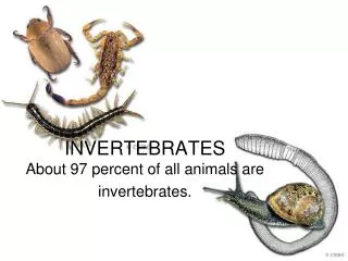
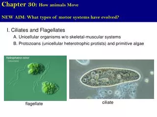
Chapter 30: How animals Move. NEW AIM: What types of motor systems have evolved?. I. Ciliates and Flagellates. A. Unicellular organisms w/o skeletal-muscular systems. B. Protozoans (unicellular heterotrophic protists) and primitive algae. ciliate. flagellate. Chapter 30: How animals Move.

E N D
Chapter 30: How animals Move NEW AIM: What types of motor systems have evolved? I. Ciliates and Flagellates A. Unicellular organisms w/o skeletal-muscular systems B. Protozoans (unicellular heterotrophic protists) and primitive algae ciliate flagellate
Chapter 30: How animals Move NEW AIM: What types of motor systems have evolved? I. Ciliates and Flagellates A. Unicellular organisms w/o skeletal-muscular systems B. Protozoans (unicellular heterotrophic protists) and primitive algae Fig. 4.18 (9 + 2 arrangement)
Chapter 30: How animals Move NEW AIM: What types of motor systems have evolved? II. Pseudopodia A. Used by amoeba to move B. Cell extensions (no microtubules) pseudopod
Chapter 30: How animals Move NEW AIM: What types of motor systems have evolved? IIB. Chemotaxis vs. Phototaxis A. Chemotaxis – process by which a cell directs their movement depending on a chemical in the environment – taxis = to move (hence the word taxi). Ex. 1. Movement of sperm towards the egg (egg secretes chemicals that sperm are attracted to); 2.Movement of macrophages to a site of bacterial infection (broken cells release a chemical attractant) 3. Movement of bacteria to a high concentration of glucose These are all examples of positive chemotaxis (move towards the chemical) There can also be negative chemotaxis (move away from the chemical).
Chapter 30: How animals Move NEW AIM: What types of motor systems have evolved? IIB. Chemotaxis vs. Phototaxis b. Phototaxis – process by which an entire organism directs their movement depending the stimulus of light (this is NOT a plant moving towards light, which is called phototropism. Ex. 1. Algal cell moves toward light (positive phototaxis) Ex. 2. Moths or fruit flies attracted to light Movement
Chapter 30: How animals Move NEW AIM: What types of motor systems have evolved? http://www.biology.ualberta.ca/facilities/multimedia/uploads/zoology/oligochaete.swf III. Hydrostatic skeletons A. Fluid held under pressure in a closed body compartment Fig. 30.1 B. Helps protect other body parts (cushion from shocks) C. Gives body shape D. Gives support for muscle action E. Cnidarians (hydra) and annelids (earthworms) (setae) Fig. 18.7 Earthworms crawl by peristalsis Each segment expands and contracts independently
Chapter 30: How animals Move NEW AIM: What types of motor systems have evolved? III. Hydrostatic skeletons A. Fluid held under pressure in a closed body compartment B. Helps protect other body parts (cushion from shocks) C. Gives body shape Fig. 30.2 D. Gives support for muscle action E. Cnidarians (hydra) and annelids (earthworms) Hydrostatic skeleton of hydra in 2 stages
Chapter 30: How animals Move NEW AIM: What types of motor systems have evolved? IV. Exoskeleton A. Hard external skeleton covering all the muscles and organs of some invertebrates B. Made of chitin in Arthropods C. Calcium carbonate in mollusks Chitin is a polysaccharide of N-acetylglucosamine (glucose with an acetyl group) D. Protection E. Limits growth (periodic molting and deposition of new exoskeleton necessary) Fig. 30.2 F. Muscles attach to inside of exoskeleton JOINTED APPENDAGES
Chapter 30: How animals Move NEW AIM: What types of motor systems have evolved? V. Endoskeleton A. Hard or leathery support tissue situated AMONG the soft tissues of animals B. Sponges (Porifera) i. Reinforce by tough protein fibers or hard calium salts/silica called spicules Spongin - fibrous protein Calcium salts A rigid sponges thanks to spicules
Chapter 30: How animals Move NEW AIM: What types of motor systems have evolved? V. Endoskeleton A. Hard or leathery support tissue situated AMONG the soft tissues of animals C. Echinoderms (sea stars, sea urchins, etc…) i. Have hard plates beneath skin (the spikes are not an exoskeleton) Sea Urchin Dead sea urchin (endoskeleton)
Chapter 30: How animals Move NEW AIM: What types of motor systems have evolved? V. Endoskeleton D. Vertebrates i. Site of muscle attachment - permits movement when muscle contracts bringing bones closer together ii. Protection and overall support - bones enclose vital organs - Ex. Rib cage surrounds thoracic organs (heart and lungs) - Ex. Skull and vertebral column surround brain and spinal cord iii. Contains both cartilage and bone - Both connective tissue
Chapter 30: How animals Move NEW AIM: What types of motor systems have evolved? V. Endoskeleton D. Vertebrates iv. cartilage - firm, but flexible - makes up skeletons of lower vertebrates (rays and sharks) - principle component of embryonic skeletons in higher vertebrates - NO blood vessels (avascular) or nerves - takes a longer time to heal than bone Fig. 30.2E Amphibian endoskeleton (cartilage in blue)
Chapter 30: How animals Move NEW AIM: What types of motor systems have evolved? V. Endoskeleton D. Vertebrates iv. cartilage - Location - articular surface of the bones (sites where 2 bones contact each other), the rib cage, the ear, the nose, the bronchial tubes/trachea and the intervertebral discs. Cartilage in yellow
Chapter 30: How animals Move NEW AIM: What types of motor systems have evolved? V. Endoskeleton D. Vertebrates v. bone - skeleton of mature higher vertebrates - composed of calcium phosphate salts and strands of collagen protein - cells in bone 1. Osteoblasts - build bone (bbb) 2. Osteoclasts - break down bone You should know which hormones interact with each cell type… - Osteoporosis - imbalance between osteoblasts and osteoclasts leading to weak bones
Chapter 30: How animals Move NEW AIM: What types of motor systems have evolved? V. Endoskeleton D. Vertebrates v. bone - spongy vs. compact bone - yellow marrow vs. red marrow Fig. 30.5 Fig. 30.3
Chapter 30: How animals Move NEW AIM: What types of motor systems have evolved? Each osteon consists of concentric layers, or lamellae, of compact bone tissue that surround a central canal, the Haversian canal through which blood vessels and nerves run. V. Endoskeleton D. Vertebrates v. bone Lacuna = the region where osteocytes reside. - highly vascular unlike cartilage Fig. 30.5
Chapter 30: How animals Move NEW AIM: What types of motor systems have evolved? Each osteon consists of concentric layers, or lamellae, of compact bone tissue that surround a central canal, the Haversian canal through which blood vessels and nerves run. V. Endoskeleton D. Vertebrates v. bone Lacuna = the region where osteocytes reside. - highly vascular unlike cartilage Fig. 30.5
Chapter 30: How animals Move NEW AIM: What types of motor systems have evolved? V. Endoskeleton D. Vertebrates v. bone - connected at joints - can be movable (your elbow) or immovable (bones of the skull) Fig. 30.3
Chapter 30: How animals Move NEW AIM: What types of motor systems have evolved? V. Endoskeleton D. Vertebrates v. bone - axial (blue - skull, ribs, vertebrae) vs. appendicular (yellow - appendages) skeleton Fig. 30.3
Chapter 30: How animals Move NEW AIM: What types of motor systems have evolved? VI. Muscle contraction and movement A. The skeleton and muscles interact in movement B. Muscle system is an EFFECTOR of the nervous system C. MUSCLES CAN ONLY CONTRACT (SHORTEN) Origin of bicep D. Insertion of a muscle i. Portion attached to bone that moves Insertion of bicep E. The ORIGIN is the attachment to the non-moving bone Origin of tricep Insertion of tricep Fig. 30.7
Chapter 30: How animals Move NEW AIM: What types of motor systems have evolved? VI. Muscle contraction and movement E. Extensor i. Muscle that extends or straightens the bones at a joint Ex. Tricep is an extensor - it contracts and straightens arm at elbow F. Flexor i. Muscle that bends a joint to an acute angle Ex. Bicep is a flexor - it contracts and bends arm at elbow Flexor Bicep and Tricep are antagonistic muscles ALL animals have pairs of antagonistic muscles Extensor Fig. 30.7
Chapter 30: How animals Move NEW AIM: What types of motor systems have evolved? VI. Muscle contraction and movement G. Tendons (dense connective tissue) i. Connect muscles to bones Ex. Achilles tendon Fig. 30.7
Chapter 30: How animals Move NEW AIM: What types of motor systems have evolved? VI. Muscle contraction and movement H. Ligaments i. Connect bones to bones
Chapter 30: How animals Move NEW AIM: What types of motor systems have evolved? VI. Muscle contraction and movement I. Three types of muscles i. smooth ii. cardiac iii. skeletal - movement caused by CONTRACTION in ALL 3 types - Contraction caused by sliding of actin and myosin filaments past each other inside cells…
Chapter 30: How animals Move NEW AIM: What types of motor systems have evolved? VI. Muscle contraction and movement I. Three types of muscles i. Smooth muscle a. involuntary muscles (autonomic NS) in arteries and veins, gastrointestinal tract, bladder, uterus b. nonstriated - simply means that actin and myosin do not have clear organized arrays c. smooth muscle cells connected by gap junctions in tissues (allow action potential to pass from one cell to next) - electrical synapse d. Single nucleus per cell
Chapter 30: How animals Move NEW AIM: What types of motor systems have evolved? VI. Muscle contraction and movement I. Three types of muscles ii. Cardiac muscle a. Single nucleus per cell b. striated - actin and myosin have clear organized arrays c. connected by gap junctions in tissues (allow action potential to pass from one cell to next) - electrical synapse d. Involuntary (autonomic NS)
Chapter 30: How animals Move NEW AIM: What types of motor systems have evolved? VI. Muscle contraction and movement I. Three types of muscles iii. Skeletal muscle a. Voluntary (intentional physical movement; somatic NS) b. Muscle cell = single, large, multinucleated fiber c. striated - actin and myosin have clear organized arrays d. Stimulated by nerves at neuromuscular synapses e. Action potential in cell stimulates calcium release into cytoplasm, which in turn causes contraction
Chapter 30: How animals Move AIM: How do muscle fibers contract? VIII. Neuromuscular junction Fig. 30.10
Chapter 30: How animals Move AIM: How do muscle fibers contract? IX. How does a motor neuron make a muscle fiber contract? Fig. 30.10 1. Action potential (AP) reaches synaptic knob 2. Acetylcholine released into synaptic cleft 3. Sodium moves through muscle fiber (just like a neuron) 4. AP travels along T-tubules (membranous tubules that fold in through cells) deep into the fiber Neuromuscular junction video on website
Chapter 30: How animals Move AIM: How do muscle fibers contract? IX. How does a motor neuron make a muscle fiber contract? 1. Action potential (AP) reaches synaptic knob 2. Acetylcholine released into synaptic cleft 3. Sodium moves through muscle fiber (just like a neuron) 4. AP travels along T-tubules (membranous tubules that fold in through cells) deep into the fiber 5. AP causes Ca++ to be released from sarcoplasmic reticulum (SR = ER) of fiber into cytoplasm Muscle action potential video on website Fig. 30.10
Chapter 30: How animals Move NEW AIM: How do muscle fibers contract? Fig. 30.8 VII. Muscle contraction A. Skeletal muscle i. Muscle composed of bundles of fibers (cells) ii. Muscle fibers (cells) contain numerous myofibril (contractile protein structures) - striation = alternating light and dark band of myofibrils iii. Sarcomere - repeating unit of the myofibril (region b/w two Z lines) – you see sarco- you think muscle - thin filament: two strands of actin polymers and one strand of regulatory protein - thick filament: staggered array of multiple myosins - dark band vs. light band
Chapter 30: How animals Move NEW AIM: How do muscle fibers contract? Sarcomere contraction video on website VII. Muscle contraction B. Sliding-filament model Fig. 30.9
Chapter 30: How animals Move AIM: How do muscle fibers contract? VII. Muscle contraction B. Sliding-filament model 1. ATP binds to myosin head (causes detachment from actin) 2. ATP hydrolyzes to ADP and Pi - energy used ratchet back the head - head is now in an unstable (high energy) state 3. Head binds to actin 4. ADP and Pi are released resulting in the power stroke 5. ATP binds, head releases, repeat again, but grab the next actin closer to Z-line Sliding filament video on website Fig. 30.9
Chapter 30: How animals Move AIM: How do muscle fibers contract? VII. Muscle contraction B. Sliding-filament model 1. ATP binds to myosin head (causes detachment from actin) 2. ATP hydrolyzes to ADP and Pi - energy used ratchet back the head - head is now in an unstable (high energy) state 3. Head binds to actin 4. ADP and Pi are released resulting in the power stroke 5. ATP binds, head releases, repeat again, but grab the next actin closer to Z-line Aside: Rigor Mortis – when an animal dies, it becomes stiff (hence why we call dead people stiffs). This is because ATP is needed to release the myosin head from the actin filaments. No ATP, no release, muscle can’t relax. Fig. 30.8
Chapter 30: How animals Move AIM: How do muscle fibers contract? IX. How does a motor neuron make a muscle fiber contract? 6. Myosin binding sites on actin usually blocked by regulatory strand (troponin and tropomyosin) 7. Ca++ binds to part of regulatory strand (troponin) of thin filament, which causes tropomyosin to move off myosin binding site so myosin can bind. Muscle action potential video on website Fig. 30.10
Chapter 30: How animals Move AIM: How do muscle fibers contract? IX. How does a motor neuron make a muscle fiber contract? http://www.tvermilye.com/pmwiki/pmwiki.php?n=Animation.Video12 Fig. 30.10
Chapter 30: How animals Move AIM: How do muscle fibers contract? X. Vocabulary for a skeletal muscle cell A. Sarcolemma: plasma membrane B. Sarcoplasmic reticulum (SR): endoplasmic reticulum C. Sarcomere: single unit of the myofibril D. Sarcoplasm: cytoplasm
Chapter 30: How animals Move AIM: How do muscle fibers contract? XI. Malfunctions A. Arthritis - inflammation of joints causing swelling and severe pain - causes: autoimmune, infection (septic), gouty arthritis, etc… B. Tendonitis - inflammation of tendon usually at site of attachment to bone caused by physical stress and irritation (common in athletes)
EXTRAS I. Heart Attack: A. Coronary Thrombosis B. Angina pectoris - narrowing of coronary arteries resulting in an inadequate supply of blood (oxygen) to heart muscle and intense pain in chest/shoulder/arm (referred pain). - typically caused by arthrosclerosis
EXTRAS II. oxyhemoglobin A. Hemoglobin with oxygen bound III. Bronchitis A. Inflammation of bronchi, caused by bacteria, virus or other irritant (i.g. tobacco smoke)
EXTRAS IV. Asthma - chronic disease where airways constrict, become inflamed, and are lined with excessive mucus. - triggered by exposure to an allergen, tobacco smoke, cold or warm air, perfume, pet dander, moist air, exercise or exertion, emotional stress. - genetic / environmental - allergic response
EXTRAS IV. Asthma Albuterol (Salbutamol) - typical used in inhalers - stimulates -2 adrenergic (adrenalin) receptors, which relaxes airway smooth muscle
EXTRAS V. CO2 carried mostly in blood as bicarbonate (carbonic acid), which acts as a buffer in the blood (pH 7.4) VI. Habits - acquired by repetition, which established pathways for nerved impulse transmission, which permit rapid automatic responses to various stimuli. - performed without thinking - DO NOT confuse with habituation
EXTRAS VII. Filament
EXTRAS VIII. Photolysis
EXTRAS VIII. Enzyme/Substrate relationship
EXTRAS IX. Enzyme/Temperature and Enzyme/pH Relationship