Download
1 / 106
1.08k likes | 1.4k Views
Duchenne muscular dystrophy (DMD) Richard C. Arceo, M. D. DESCRIPTION Duchenne muscular dystrophy (DMD) is a severe recessive X-linked form of muscular dystrophy characterized by rapid progression of muscle degeneration , eventually leading to loss of ambulation and death.

E N D
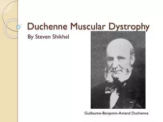
Duchenne muscular dystrophy (DMD) Richard C. Arceo, M. D.
DESCRIPTION Duchenne muscular dystrophy(DMD) is a severe recessive X-linked form of muscular dystrophy characterized by rapid progression ofmuscle degeneration, eventually leading to loss of ambulation and death.
Incidence/Prevalence • Although reliable prevalence data are lacking, the prevalence of DMD is generally estimated at 1:3,500 live male births (Emery 1991). • The birth prevalence of DMD in northern England isone in 5,618 live male births.
Pathogenesis • DMD is caused by mutations in the dystrophin gene which is the largest human gene, spanning 2,200 kb on the X chromosome and occupying roughly 0.1% of the genome. • The gene is composed of 79 exonsand 8 tissue-specific promoters [Koenig et al., 1987]. • The primary transcript measures about 2,400 kilobases and takes 16 hours to transcribe, the mature mRNA measures 14.0 kilobases. • The 79 exons code for a protein of over 3500 amino acid residues.
Dystrophin is a rod-shaped cytoplasmic protein, and a vital part of a protein complex that connects the cytoskeleton of a muscle fiber to the surrounding extracellular matrix through the cell membrane. • Dystrophin provides structural stability to the Dystroglycan complex (DGC), located on the cell membrane
Abnormal gene product • Mutations will lead to lack of dystrophin expression causing DMD, whereas those that lead to abnormal quality or quantity of dystrophin lead to BMD.
Much investigative work determined that dystrophin is involve in the release of calcium from the sarcoplasmic reticulum in muscle fibers. The lack of dystrophin causes calcium to leak into the cell, which promotes the action of an enzyme that dissolves muscle fibers. When the body attempts to repair the tissue, fibrous tissue forms, and this cuts off the blood supply so that more and more cells die.
CLINICAL FEATURES SKELETAL MUSCLE • The most distinctive feature of Duchenne muscular dystrophy is a progressive proximal muscular dystrophy with characteristic pseudohypertrophy of the calves
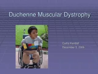
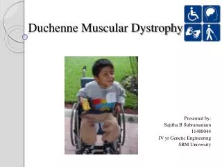
The first symptoms of DMD appear duringpreschool years. The disorder affects thelegs first. A boy hastrouble walking and maintaining balance. In most cases, he begins walking three to six months later than average.
As his muscles begin to weaken, he may change the way he walks. He places his legs farther apart in order to maintain balance. Walking this way produces a waddling effect that is characteristic of DMD.
Contracturesusually begin at about the age of five or six. This forces a boy to walk on his tiptoes. Balance becomes more of a problem. As a result, falls and broken bones become common at this age.
By the age of nine or ten, a boy with DMD might not be able to climb stairs Or stand by himself. By age 10, braces may be required to aid in walking but most patients are wheelchair dependent by age 12.
Early common sign of muscular dystrophy To get up from the ground, the child ‘walks up' his thighs with his hands. This is mainly because of weak thigh muscles.
The Baskaran familySouth East of England Jamie Oliver
May develop a severe curve of the spine. • Heart and breathing muscles also get weak. • Child usually dies before age 20 from heart failure or pneumonia.
NERVOUS SYSTEM • Mental retardation of mild degree is a pleiotropic effect of the Duchenne gene (Zellweger and Niedermeyer, 1965) • As indicated later, the finding of dystrophin mRNA in brain may bear a relationship to the mental retardation in DMD patients. • In 50 DMD patients with a mean age of 11.1 years (range 3.5 to 20.3), Bresolin et al. (1994) found that31% had a Wechsler full intelligence quotient (FIQ) lower than 75 and that only 24% had appropriate IQ levels by this index
Bushby et al. (1995) studied 74 boys with DMD, 18% of which had a full scale IQ of below 70. The authors found no significant IQ difference between the patients with promoter deletions and those without, nor did they find a relationship between the length of the deletion and full scale IQ. They found, however, that boys with distal deletions were more likely to be mentally retarded than were those with proximal deletions
CARDIAC MUSCLE • Myocardial involvement appeared in a high percentage of DMD patients by about 6 years of age; it was present in 95% of cases by the last years of life. (Nigro et al., 1983). • Mirabella et al. (1993) noted that electrocardiographicabnormalities had been reported in 6.6 to 16.4% of DMD heterozygous females and that in one carrier female severe cardiomyopathy had been described in association with muscle weakness. • They reported 2 carriers with dilated cardiomyopathy and increased serum CK but no symptoms of muscle weakness. Heart biopsies in both patients showed absence of dystrophin in many muscle fibers
SMOOTH MUSCLE • Noting that in DMD functional impairment of smooth muscle in the gastrointestinal tract can cause acute gastric dilatation and intestinal pseudoobstruction that may be fatal, Barohn et al. (1988) studied gastric emptying in 11 patients with DMD. • Strikingly delayed gastric emptying times were observed.
Boland et al. (1996) studied a retrospective cohort of 33 male patients born between 1953 and 1983. The mean age at DMD diagnosis was 4.6 years;wheelchair dependency had a median age of 10 years; cardiac muscle failure developed in 15% of patients with a median age of 21.5 years; smooth muscle dysfunction in the digestive or urinary tract occurred in 21% and 6% of the patients, respectively, at a median age of 15 years. In this cohort, deathoccurred at a median age of 17 years.
Diagnosis 1. Serum creatine phosphokinase (CK) concentration
2. Electromyography (EMG) is useful in distinguishing a myopatic process from a neurogenic disorder. This is done by demonstrating short-duration, low-amplitude, polyphasic, rapidly recruited motor unit potentials.
3. Skeletal muscle biopsy • Muscle histology early in the disease shows nonspecific dystrophic changes, including variation in fiber size, foci of necrosis and regeneration, hyalinization, and, later in the disease, deposition of fat and connective tissue.
Findings in the Dystrophin Protein from Skeletal Muscle Biopsy
Molecular Genetic Testing • Gene: DMD is the only gene known to be associated with DMD • Clinical testing: Deletion/duplication Analysis 1. Multiplex PCR [Multicenter Study Group 1992], 2.Southern blotting [Darras et al 1988], and 3. FISH (with probes covering DMD exons 3-6, 8, 12, 13, 17, 19, 32-34, 43-48, 50, 51, and 60) can be used to detect deletions, which account for approximately 65% of mutations in individuals with DMD. Approximately 98% of deletions are detectable by these methodologies.
Southern blotting and quantitative PCR analysis can be used to detect duplications. Duplications may lead to in-frame or out-of-frame transcripts and account for the disease-causing mutations in approximately 6%-10% of males with DMD or BMD. In one study [Galvagni et al 1994], duplications were detected in 8.18% of individuals with DMD.In a series of individuals already screened for deletions and point mutations, duplications were detected in 87% of cases [White et al 2006].
New testing methods including single-condition amplification internal primer sequencing (SCAIP) [Flanigan et al 2003] and denaturing gradient gel electrophoresis (DGGE)-based whole-gene mutation scanning [Hofstra et al 2004] aim at detecting the remaining 30%-35% of the DMD mutations in a semiautomatic, rapid, accurate, and economical fashion. • A muscle biopsy-based diagnostic approach was developed and optimized to increase the mutation detection frequency to nearly 100% [Deburgrave et al 2007].
To date, 501 deletions, 8 duplications, and 989 point mutations have been documented in the dystrophin gene (Leiden muscular dystrophy database; www.dmd.nl). • 5 exons commonly deleted in deletion-type Duchenne muscular dystrophy (DMD). • The five DMD gene exons (17, 19, 44, 45 and 48) can be analysed in separate duplex PCR reactions
The current methodologies used for detecting mutations in the dystrophin gene include multiplex PCR, Southern blotting [Stockley et al., 2006], multiplex ligation-dependent probe amplification (MLPA) [Gatta et al., 2005; Janssen et al., 2005; Schwartz and Duno, 2004], detection of virtually all mutations-SSCP (DOVAM- S) [Buzin et al., 2000, 2005; Liu et al., 1999], denaturing high-performance liquid chromatography (DHPLC) [Bennett et al., 2001], single condition amplification/internal primer sequencing (SCAIP) [Flanigan et al., 2003], and Sanger sequencing [Hamed and Hoffman, 2006; Stockley et al., 2006]. • HUMAN MUTATION 0,1^9,2008
Signs and Symptoms in Carriers of Duchenne and Becker Muscular Dystrophy DMD Carriers BMD Carriers • None 76% 81% • Muscle weakness19% 14% • Myalgia/cramps 5% 5% • Left ventricle dilation19% 16% • Dilated cardiomyopathy8% 0 From Hoogerwaard et al [1999b)
Carrier Testing • A reliable and simple method based on quantitative real-time PCR detects deletions/duplications in 100% of DMD/BMD carriers [Joncourt et al 2004]. • Carrier testing for deletions may also be performed by FISH [Voskova-Goldman et al 1997]. • Carrier testing for point mutations may be performed by sequence analysis.
Genotype-Phenotype Correlations • In males with DMD, phenotypes are best correlated with the degree of expression of dystrophin, which is largely determined by the reading frame of the spliced message obtained from the deleted allele [Monaco et al 1988, Koenig et al 1989]. • Very large deletions may lead to absence of dystrophin expression. • Mutations that disrupt the reading frame include stop mutations, some splicing mutations, and deletions or duplications. • They produce a severely truncated dystrophin protein molecule that is degraded, leading to the more severe DMD phenotype.
Data suggest that dystrophin deletions involving the brain distal isoform Dp140 are associated with intellectual impairment [Felisari et al 2000
Testing Strategy Establishing the diagnosis of DMD: • For individuals with clinical findings suggesting a dystrophinopathy and an elevated serum CK concentration, the first step in diagnosis is molecular genetic testing of the DMD gene: • If a disease-causing mutation is identified, the diagnosis is established; • If no DMD disease-causing mutation is identified, skeletal muscle biopsy of individuals with suspected DMD is warranted for western blot and immunohistochemistry studies of dystrophin.
Management Evaluations Following Initial Diagnosis To establish the extent of disease in an individual diagnosed with a dystrophinopathy, the following evaluations are recommended: • Physical therapy assessment • Developmental evaluation before entering elementary school for the purpose of designing an individualized educational plan, as necessary • If the individual is older than age ten years at diagnosis, evaluation for cardiomyopathy by electrocardiography, chest radiography, cardiac echocardiography, pulmonary function studies, and/or MRI [Towbin 2003]
Medications • Prednisone.Studies have shown that prednisone improves the strength and function of individuals with DMD. It is hypothesized that prednisone has a stabilizing effect on membranes and perhaps an anti-inflammatory effect. Whether the improvement is the result of an immunosuppressive effect remains unclear, as individuals treated with azathioprine did not have a beneficial effect.
In a randomized double-blind six-month trial, prednisone administered at a dose of either 0.75 mg/kg/day or 1.5 mg/kg/day increased strength and reduced the rate of decline in males with DMD [Mendell et al 1989]. • The improvement begins within ten days of starting the treatment, requires a single dose of 0.75 mg/kg/day of prednisone for maximal improvement, reaches a plateau after three months, and can be sustained for as long as three years in those children maintained on doses of 0.5 and 0.6 mg/kg/day [Fenichel et al 1991]. • One open-label study suggested that therapy with prednisone could prolong ambulation by two years. • Side effects include weight gain (>20% of baseline) (40%), hypertension, behavioral changes, growth retardation, cushingoid appearance (50%), and cataracts [Mendell et al 1989, Griggs et al 1993].
Pulmonary: • Baseline pulmonary function testing before confinement to a wheelchair (usually age ~9-10 years) • Evaluation by a pediatric pulmonologist twice yearly after any one of the following: confinement to a wheelchair, reduction in vital capacity below 80% predicted, and/or age 12 years [Finder et al 2004]
Deflazacort: • Deflazacort, a synthetic derivative of prednisolone used in Europe but not currently available in the US, is thought to have fewer side effects than prednisone, particularly with regard to weight gain [Angelini 2007]. • A larger study comparing deflazacort to prednisone, carried out in Europe, showed that the two medications were similarly or equally effective in slowing the decline of muscle strength in DMD. • Another European multicenter, double-blind, randomized trial of deflazacort versus prednisone in DMD showed equal efficacy in improving motor function and functional performance [Bonifati et al 2000]. • A more recent study of deflazacort treatment showed efficacy in preserving pulmonary function as well as gross motor function [Biggar et al 2006].
In a comparison of two different protocols of deflazacort treatment in DMD, a 0.9-mg/kg/day dose was more effective than a dose of 0.6 mg/kg/day for the first 20 days of the month and no deflazacort for the remainder of the month [Biggar et al 2004]; 30% of children on the highest dose developed asymptomatic cataracts that required no treatment. • A systematic review and meta-analysis of 15 studies showed that deflazacort improves strength and motor function more than placebo; whether it has a benefit over prednisone on similar outcomes remains unclear [Campbell & Jacob 2003].
Therapies Under Investigation • Aminoglycosides. Up to 15% of individuals with DMD exhibit the gene mutation known as a premature stop codon. • Suppression of stop codons has been demonstrated with aminoglycoside treatment of cultured cells; the treatment creates misreading of RNA and thereby allows alternative amino acids to be inserted at the site of the mutated stop codon. • In the mdx mouse, in vivo gentamicin therapy resulted in dystrophin expression at 10%-20% of that detected in normal muscle [Barton-Davis et al 1999], a level that provided some degree of functional protection against contraction-induced damage.
Aminoglycoside therapy has been suggested as an alternative to gene therapy but could be aimed only at individuals with premature stop codons. In a preliminary study in which gentamicin (7.5 mg/kg/day) was administered to four individuals for two weeks, full-length dystrophin did not appear in the muscles of the treated individuals [Wagner et al 2001]. Some authors, unable to reproduce the results previously published for the mouse model of DMD, have called for more preclinical investigation of this potential therapy [Dunant et al 2003]. In an in vitro study [Kimura et al 2005], dystrophin expression was detected in myotubes of males with DMD using gentamicin; however, the treatment was more effective in persons with the nonsense mutation TGA than TAA or TAG.
PTC124is a new, orally administered non-antibiotic drug that appears to promote ribosomal read-through of nonsense (stop) mutations. Preclinical efficacy studies in mdx mice have yielded encouraging results [Barton et al 2005, Welch et al 2007]. A Phase I multiple-dose safety trial is ongoing [Hirawat et al 2005]. • Morpholino antisense oligonucleotides mediate exon skipping [Aartsma-Rus et al 2006a] and have improved the mdx mouse model of DMD [Wilton & Fletcher 2005, Alter et al 2006].
Oxandrolone, an anabolic (androgenic) steroid with a powerful anabolic effect on skeletal muscle myosin synthesis [Balagopal et al 2006], was shown in a pilot study to have effects similar to prednisone, with fewer side effects [Fenichel et al 1997]. • A randomized, prospective, controlled trial showed that oxandrolone did not produce a significant change in the average manual muscle strength score of males with DMD, as compared with placebo; however, the mean change in quantitative muscle strength was significant [Fenichel et al 2001]. • The investigators conducting this study felt that oxandrolone may be useful before initiating therapy with corticosteroid because it is safe in the short term, accelerates linear growth, and may be beneficial in slowing the progression of weakness. • However, the long-term effects of oxandrolone in the treatment of DMD have not been studied.