Download
1 / 27
270 likes | 344 Views
Understanding multi - cellular systems. Jin Chen CSE891-001 2012 Fall. EXAMPLE.

E N D
Understanding multi-cellular systems Jin Chen CSE891-001 2012 Fall
EXAMPLE Multi-cellular signaling network of 169 genes/proteins regulated during the first 4 hours of EGFR activation in human mammary cells. Results were obtained by integrating microarray, proteomic and Western blot data in PNNL. http://www.sysbio.org/sysbio/multicellular/index.stm
Why multi-cellular systems? • Understanding multi-cellular systems is an imperative element for adapting systems biology studies to medical applications • Example: understand host-pathogen interactions • How cells on the defensive front lines interact with an invader • How different host cell types involved in the inflammatory cascade triggered by an infection interact with one another • Studying signaling among different cell types in a single organism is important for understanding diseases and the processes involved in disease, such as metastasis. http://www.sysbio.org/sysbio/multicellular/index.stm
Strategies to study multi-cellular networks • Expand the capabilities of analytical and computational tools • Coupling the development of both modeling and experimental approaches • Reduce the enormous complexity of biological organisms to simplest terms http://www.sysbio.org/sysbio/multicellular/index.stm
Rules to follow • The usefulness of high-throughput data can be greatly increased by integrating multiple data types that are obtained in parallel on the same system • Signaling networks can be accurately modeled as a series of functional modules instead of networks of individual interactions http://www.sysbio.org/sysbio/multicellular/index.stm
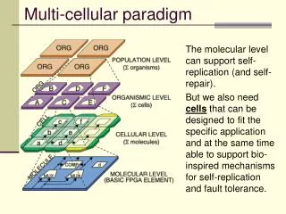
PNNL’s functional module approach • Reduce the network complexity by describing them in terms of functional modules, rather than individual molecular reactions - follow the flow of information among multiple cell types to connect molecular events to overall system behavior • A top-down perspective • Consider a biological system as an input-output system. • Collect baseline, multidimensional data on the overall system. • Analyze the data in the context of the literature and current databases to identify functional modules. • Construct a high-level model based on tentatively identified functional modules. • Make testable predictions of outputs in response to specific inputs and test those predictions with experiments specifically designed to enable step 6. • Use differences between predictions and experiments to refine the original model, adding more modules when necessary. • Repeat 5
MetaCore • MetaCoreis an integrated knowledge database and software suite for pathway analysis of experimental data and gene lists. • MetaCoreis based on a proprietary manually curated database of human protein-protein, protein-DNA and protein compound interactions, metabolic and signaling pathways for human, mouse and rat, supported by proprietary ontologies and controlled vocabulary. • The analytical package includes tools for search, data visualization, mapping and exchange, biological networks and interactome. • MetaRodent • MetaLink • MetaSearch http://www.genego.com
Multi-cellular differentiation • Understanding the processes involved in multi-cellular pattern formation is a central problem of developmental biology. • Defining suitable computational techniques for development modeling, able to perform in silico simulation experiments, is an open and challenging problem. 2009 Bonzanni N et al. Bioinformatics 2009;25:2049-2056
Background • Many efforts have been undertaken to elucidate how cells are able to coordinate different and sometimes conflicting signals, producing a precise phenotype during the animal organogenesis • C. elegansvulva developmentprovides an elegant and relatively well-charted model to study how multiple pathways, in multiple cells, interact to produce developmental patterns. VPC
Background • The first diagrammatic model, describing the regulatory network underlying VPC determination, was proposed by Sternberg and Horvitz (1989). Since then, global understanding of the biological network has improved greatly. • The first computational model, proposed by Kamet al.(2003), combined multiple experimental ‘scenarios’ from Sternberg and Horvitz (1986) into a single model, using Live Sequence Charts (LSCs). • Afterwards, in two landmark papers, Fisher et al., 2005, 2007) suggested two state-based mechanistic models. • Two other insightful models of C.elegansvulval development have been published. Giurumescuet al. (2006) proposed a partial model based on ODEs, while Sun and Hong (2007) developed a model based on automatically learned dynamic Bayesian networks with discrete states. • Lietal. (2009) recently modeled part of C.elegansvulval development using hybrid functional Petri nets with extensions.
Petri nets • Apply a discrete, non-deterministic Petri nets-based model to C.elegansvulval development. • Petri nets are a convenient formalism to represent biological networks. This formalism models process synchronization, asynchronous events, conflicts and in general concurrent systems in a natural way. • Petri nets offer direct insights into causal relationships, and allow a graphical visualization that resembles the diagrams used to describe biological knowledge.
Petri net A Petri net is a directed bipartite graph, in which the nodes represent transitions (bars) and places (circles). The directed arcs describe which places are pre- and/or post-conditions for which transitions occurs. Some sourcesstate that Petri nets were invented in August 1939 by Carl Adam Petri — at the age of 13 — for the purpose of describing chemical processes. He documented the Petri net in 1962 as part of his dissertation http://en.wikipedia.org/wiki/Petri_net
Petri net • Places in a Petri net contain a discrete number of marks called tokens. • Any distribution of tokens over the places will represent a configuration of the net called a marking. • In an abstract sense relating to a Petri net diagram, a transition of a Petri net may fire whenever there are sufficient tokens at the start of all input arcs • When it fires, it consumes these tokens, and places tokens at the end of all output arcs. A firing is atomic, i.e., a single non-interruptible step.
Model Design • Aim: to mimic the underlying biological mechanisms as much as possible, and not only to reproduce the expected phenotype according to a specific set of mutations. • To achieve this, a principle of maximal parallelism is applied, and is bounded execution with overshooting. • Using this simple framework, we can identify different modules, each corresponding to different biological functions. • Thus, combining functional modules into cells, and joining such cells together, we iteratively developed the whole network.
Model Design • Focus on preserving the simplicity of the formalism, and develop an execution semantics which resembles biology • Places = genes, protein species and complexes • Transitions = biological processes • Firing of a transition is execution of a process, e.g. consuming substrates or creating products • Number of tokens is interpreted in two ways. • For genes as a Boolean value, 0 means not present and 1 present. • For proteins, we use abstract concentration levels 0−6: going from not present, via low, medium and high concentration to saturated level. • The rationale behind this approach is to abstract away from unknown absolute molecule concentration levels, as we intend to represent relative concentrations.
Model Design - Maximal parallelism • The maximal parallel execution semantics can be summarized informally as execute greedily as many transitions as possible in one step. • Definition: A maximally parallel step 𝒮 is a step that leaves no enabled transitions in the net, and in principle should be developed in such a way that it corresponds to one time step in the evolution of the biological system. • The modeler can capture relative speeds using appropriate weights on arcs. Typically, if in one time unit a protein A is produced four times more than a protein B, then the transition that captures production of A should have a weight that is four times as large as the weight of the one that captures B production.
Model Design - Maximal parallelism • Implementing a pure maximally parallel semantics requires to generate all possible partitions of tokens, and select one randomly, uniformly. However, with the growth of the network, this procedure becomes prohibitively slow. • This paper approximated it by building a maximally parallel step incrementally, selecting one transition after another, randomly, until all enabled transitions have been exhausted.
Model Design - Maximum capacity • Unrestricted production of proteins is usually not realistic, as in nature the cell would saturate with the product, and the reaction would slow down or stop. • To guarantee that the highest concentration level can be attained, we introduced bounded execution with overshooting. • Each place has a predefined maximum capacity 𝒩 = 6 • A transition can only fire if each output place holds fewer than 𝒩 tokens • Since each transition can possibly move more than one token at once into its output places, each transition can overshoot the pre-given capacity 𝒩 at most once. Therefore, the network is bounded with a finite bound k ≥ 𝒩.
Model design – example VAV-1 down-regulation by decreasing the translation rate of the gene vav-1. If mir-61 is not present, the reaction VAV-1 PRO is enabled and produces the protein. However, when mir-61 is present, the reaction VAV-1 DR is enabled and has 50% chance of firing compared with VAV-1 PRO, thus the production of VAV-1 will halve. Two connected basic modules, a gene expression and the endocytosis mediated down-regulation of LIN-12. Activation of the Ras/MAPK cascade leads to the transcription of a hitherto unknown gene that enhances the LIN-12 endocytosis.
Results • Petri net model for cell fate determination during C.elegansvulval induction. • The entire network comprises 600 nodes (places and transitions) and 1000 arcs. It includes the VPC network out of six interconnected cells as identical modules of a multi-potent cell. A separate block for the AC (producing the inductive signal) and for the hyp7 is also built. • It helps us to identify different modules that correspond to different biological functions, such as gene expression, protein activation and protein degradation.
Schematic representation of the whole system. • How the six VPCs, AC and hyp7 modules are connected. Adjacent cells are linked with each other, the hyp7 connects to all six cells, and the AC can directly influence cells P5.p, P6.p and P7.p.
Results • Multi-level network • Level 1: basic biological functions • Level 2: protein interactions • Level 3: pathways • Level 4: cells • Level 5: multi-cellular interactions Level 3
Results • Multi-level network • Level 1: basic biological functions • Level 2: protein interactions • Level 3: pathways • Level 4: cells • Level 5: multi-cellular interactions Level 4
Results • Multi-level network • Level 1: basic biological functions • Level 2: protein interactions • Level 3: pathways • Level 4: cells • Level 5: multi-cellular interactions Level 5
Results Comparison between photomicrographs of gene activity by fluorescently labeled gene products, and simulation results. (a) Photomicrographs of the graded expression of the inductive signal adapted from Yoo et al. (2004). (b) Time series plot generated by this model, showing the graded expression of the inductive signal, initially faintly present in P5.p and P6.p. Maximally parallel steps on the horizontal.