Download
1 / 27
290 likes | 495 Views
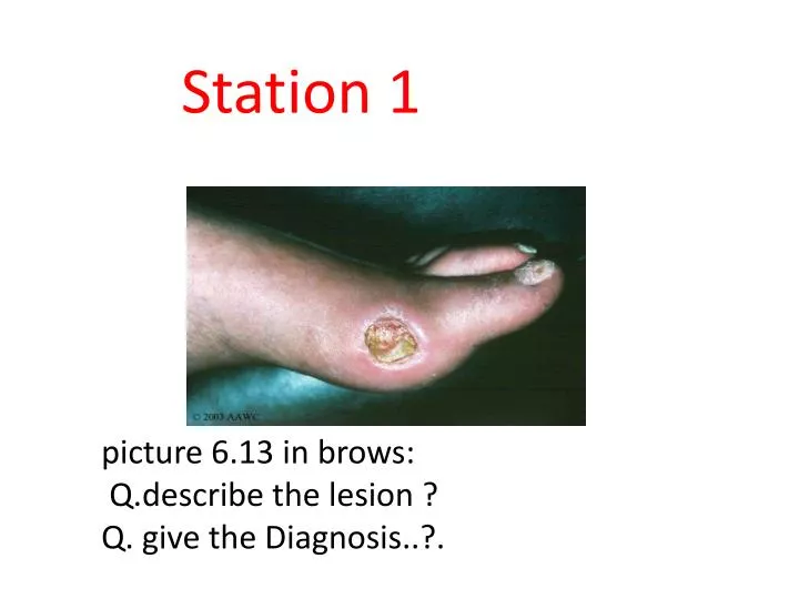
Station 1. picture 6.13 in brows : Q.describe the lesion ? Q. give the Diagnosis ..?. Describe : Round ulcer in the lateral side of the head of the first metatarsal of the left foot Measuring around __cm x __cm Regular, red floor, no discharge Edge.. Depth.. Dx : ischemic ulcer.

E N D
Station 1 picture 6.13 in brows: Q.describethe lesion ? Q. give the Diagnosis..?.
Describe : Round ulcer in the lateral side of the head of the first metatarsal of the left foot Measuring around __cm x __cm Regular, red floor, no discharge Edge.. Depth.. Dx: ischemic ulcer
Typical diabetic foot ulcer caused by high plantar pressures at the second metatarsal head.
Trophic (neuropathic) ulcer Ischemic ulcer Painful Area of repeated pressure and trauma Inadequate circulation (arterial insufficiency) Femoral & iliac art atherosclerosis Painless Area of repeated pressure and trauma Inadequate sensory nervous system Diabetic peripheral neuropathy,leprosy & 1ry neurologic abnormality
Etiology of diabetic foot Atherosclerosis Peripheral neuropathy (DM>10 yrs) Impaired immunity Charcot's or Neuropathic joint (painless & disorganized joint with abnormal weight bearing) <<go back to browse p.140 for the causes of such a deformity..
Bedridden pt Pressure areas Ambulant pt Back of the heel Lateral side of the foot Heel Ball of the foot Tips of the toes Mainly in the sole of the foot
When you examine a patient with an ulcer on their foot • EXAMINE THE CIRCULATION(pulses..) • EXAMINE THE SENSORY NERVES(light and deep touch and pain sensation) • ALSO EXAMINE THE reflexes and the motor tone & power • TEST THE URINE FOR SUGAR • Plus the whole LL examination • DON’T FORGET TO EXPOSE BOTH LIMBS
There are several signs and symptoms which should prompt the diagnosis of arterial ulcer. Some of these symptoms are: • Cool or cold skin • Reduced or absent pulse in the affected extremity • Shiny, tight, dry skin • Skin may be hairless • Toenails thickened and brittle • Ulcers small and circular in appearance • Wound edges smooth • Wound base is pale in colour • Minimal fluid drainage (unless wound is infected)
Wagner Ulcer Classification System Grade Lesion 0 No open lesions; may have deformity or cellulitis 1 Superficial diabetic ulcer (partial or full thickness) 2 Ulcer extension to ligament, tendon, joint capsule, or deep fascia without abscess or osteomyelitis 3 Deep ulcer with abscess, osteomyelitis, or joint sepsis 4 Gangrene localized to portion of forefoot or heel 5 Extensive gangrenous involvement of the entire foot
RF Don’t forget the risk factors (modifiable, non-modifiable) for the development of this type of ulcer, including Old age DM HTN smoking obesity sedentary lifestyle family history hyperhomocysteinemia hypertriglyceridemia Hyperuricemia and stress.
Invstx: Doppler US Mx: The primary goal in the Rx of diabetic foot ulcers is to obtain wound closure. Mx of the foot ulcer is largely determined by its severity (grade) and vascularity, and the presence of infection Foot elevation & relief the pressure Assess the wound daily for appearance, drainage, increase in size, signs of infection, and moistness Document wound appearance and size frequently Assess blood flow frequently Control hyperglycemia Judicious debridement of necrotic tissue & calluses Manage pain appropriately Cleanse wounds with normal saline or another non-cytotoxic cleanser Moisturize the skin of the affected limb frequently Choose the right dressing for the wound in its current state-hydrocolloids, hydrogels, foams, and calcium alginates may be acceptable choices Ensure adequate nutritional status Improve circulation, revascularization if indicated
If infected !!! Plus wt was mentioned in prev slide.. Radiograph; R/O soft tissue gas or forign body Broad spectrum abxampicillin/sulbactam or piperacillin/tazobactam Drain abscess and debride non viable tissue Aggressive wound care & whirlpool therapy Absolute non-weight bearing If infection cleared may consider revascularization DONE BY : ALHATOON AL-NAJASHI
Take Hx • When did u notice the ulcer? • What drew ur attention to it (pain, itching, etc) • What are the symptoms of the ulcer? • How did it change from its 1st appearance? • Have u had any similar ulcer on the same site or elsewhere? • What do u think is the cause of the ulcer?
Ex :Inspection • Describe : • 4s ( site, size, shape,& surface), edge, depth, discharge, surrounding tissue, state of local lymph gl • Other • webspaces: cracked, infected, ulcers, maceration • toe nails: dystrophic, in-grown, paronychia, Onychomycosis
Edge • Sloping (venous ulcer) • Punched out (neuro,arteial) • Undermined • Rolled (rodent ulcer, slow growing) • Everted (fast growing)
discharge • Serous, sanguineous, sero-sanguineous, or purulent • Quantity? • Oder?
Relation • If its close to or invading deep structures as bone, pereosteom & tendon -> osteomyelitis ?
LN • It maybe enlarged d.ttruma or infection
Palpation • For the base • The base= Floor • It consists of slough or granulation tissue (capillaries, collagen, fibroblasts, inflammatory cells) • Tendon or bone might be visible • DDX: • Poor granulation tissue + tendon + other tissue -> ischemic ulcer • Yellow-grayish leathery Slough -> Syphilitic ulcer • Bluish unhealthy granulation tissue ->TB ulcer
Palpation • Pulses • dorsalis pedis • posterior tibial • popliteal • Femoral • Temperature • use back of hand, compare shin to feet, bilaterally • temperature should decrease slightly as you get toward the toes • Other • Capillary refill
Auscultation • Bruits: femoral, popliteal
Neurological • Sensory • ↓ vibration (1st modality to loose, 128 Hz) • ↓ light touch (microfilament) • ↓ pin prick • ↓ proprioception • ↓ temperature • (loss in glove and stocking distribution) • Autonomic • ↓ sweating • dry cracked skin • Motor • intrinsic muscle wasting (clawed, hammer toes) • Pes planus, Pes cavus • Charcot joints (medial and laterial deviation at subtalar joint) • Reflexes • DTR: ↓ ankle jerk
DDX • Large A obliteration : • Atherosclerosis, Embolism • Small A obliteration • Scleroderma, burger dis, embolism, DM, Physical agents (press necrosis, radiation, trauma, electric burns)
Pt education • Daily foot inspection by the patient (or a caretaker if the patient lacks sufficient visual acuity or mobility to perform the examination) is the cornerstone of proper foot care. • Gentle cleansing with soap and water, followed by the application of topical moisturizers, helps to maintain healthy skin that can better resist breakdown and injury. • shoes for areas of inadequate support or improper fit. • Minor foot injuries and infections, such as cuts, scrapes, blisters and tinea pedis, can be unintentionally exacerbated by home remedies that impede healing. Patients should be reminded to avoid hot soaks, heating pads and harsh topical agents • Gentle cleansing of minor wounds and the application of a topical antibiotic to maintain a moist wound environment can help to prevent ulcer formation. • DONE BY : AljohrahALamer