Download
1 / 23
290 likes | 589 Views
The Eye and sight. Option A1. Structure of the Human Eye www.daviddarling.info/encyclopedia/E/eye.html. Structure of the Human Eye. Nearly spherical in shape Diameter ≈ 2.5 cm Light enters through cornea (transparent membrane) and is refracted Index of refraction for cornea is 1.37

E N D
The Eye and sight Option A1
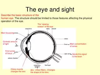
Structure of the Human Eyewww.daviddarling.info/encyclopedia/E/eye.html
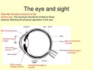
Structure of the Human Eye • Nearly spherical in shape • Diameter ≈ 2.5 cm • Light enters through cornea (transparent membrane) and is refracted • Index of refraction for cornea is 1.37 • In between cornea and eye lens is a liquid-filled chamber called aqueous humour- clear and comprised mainly of salts with index of refraction of 1.33 (almost the same as water)
Structure of the Human Eye • The aqueous humour is separated into 2 parts by the iris or coloured part of the eye. • Centre of the iris is the pupil- an aperture which allows light to enter eye lens. • Pupil can dilate i.e increase/decrease in diameter to adjust to varying intensities of light. • Eye lens is attached to ciliary muscle by ligaments and the ciliary muscle controls the curvature of the lens
Structure of the Human Eye • Light passing through lens enters second chamber filled with jelly-like substance called vitreous humour then reaches back surface of the eye, the retina • Retina is covered with light sensitive cells which record the arrival of light-two main types are rods and cones. • Light is converted into tiny electrical signals in nerve fibres attached to the rods and cones. The nerve fibres all converge to optic nerve which transmits electrical signals to brain
Structure of the Human Eye • Close to beginning of optic nerve almost on the principal axis of the eye is an area called the fovea, a spot ≈ 0.25 mm in diameter where vision is exceptionally acute • The fovea is filled with cones, each connected to a different nerve fibre unlike other places on the retina where many different cones are connected to the same fibre
Structure of the Human Eye • Distribution of rods and cones along surface of retina is not even or constant • At fovea there are lots of cones but no rods • Density of cones at fovea ≈ 150 000 mm-2 • Rods mainly found at edges of retina away from principal axis whereas concentration of cones increase closer to principal axis
Depth of Visionwww.antonine-education.co.uk/Physics_A2/optio... • A converging lens is shown below with the focal point (aka principal focus) and focal length.
Depth of Vision • Our eyes are not able to focus on two objects at two different distances from the eye. • If we focus on an object, O, far from the eye and straight ahead, so that we can see O clearly, then we will still be able to see other objects both closer and farther than O . If the furthest object that can be seen reasonably clearly is O1 and the closest is O2 , then the distance O1 O2 is called the depth of vision or depth of field
Depth of Vision • The depth of vision (dov) is the range of object distances from the eye within which objects, or points on an object , can be seen acceptably clearly. • It depends on the distance to the object- that farther the object is from the eye, the larger the dov. If the object is close to the eye, the dov is substantially reduced. If brighter light is used, dov increases since the pupil diameter decreases, and the rays of light can be focused.
Accomodation • Refers to the ability of the eye lens to change its focal length. This is done by contractions of the ciliary muscle. • When the muscle is relaxed, the ligaments are taut, and the lens is long and thin, distant objects are focused without fatigue and the eye is said to be unaccomodated. • When the muscle is contracted, the ligaments are slack and the lens is short and thick nearby objects are focused with fatigue.
Accomodation • The near point is the nearest distance which can be clearly seen without undue strain on the eye. It is ≈ 25 cm. • The far point is the furthest distance the eye can focus on clearly which is ∞ for the normal healthy eye.
Scotopic and photopic vision • Vision in which the rods are the main detectors on the incident light in the eye is called scotopic vision • Vision in which the cones are the main detectors of the incident light in the eye is called photopic vision
Colour • Perception of colour is made possible because of three types of cone cell each sensitive to either blue, green, or red light. • Refer to graph in Tsokos p. 476. • NB light of λ = 550 nm will excite only the green and red cones. In equal quantities , green and red gives yellow light so this is the colour the brain understands for this wavelength
Colour Blindnesswww.twodocs.com/color-blind-tests/total-color... • Normal will trace the blue-green line between the two X's. The majority of those with colour vision deficiencies will be unable to follow the line or will follow a line different to the normal one.
Colour Blindness • Refers to deficiency in the perception of colour. • Affects men more than women • Complete colour blindness (cb) is rare • Since colour is perceived by cone cells, cb is associated with malfunction of cone cells or insufficient numbers of one or more types of the cells. May also be due to brain/nerve damage
Colour Blindness • Most common form involves green and red cells and inability to distinguish between these two colours. • If one type of cone cell malfunctions, then the colours which can be perceived are only those made by combining colours to which the other two types of cone cell are sensitive. • If two types of cone cell malfunction, the person is completely colour blind since they can’t distinguish between any two coloured objects.
Colour Addition • Mixing three colours gives a very wide range of other colours. • If G = green, R = red, and B = blue and g,r,and b are their relative intensities, then X = gG + rR + bB gives any colour • Green, Red, and Blue are called primary colours i.e. if green, red and blue light of different intensities were shone on a white screen, the colour X would appear where the three overlap.
Colour addition • Primary colours are those which when overlapped, give a wide range of other colours. No one primary colour can be made with the other two. Adding the three primary colours gives white light. • W = B + G + R • Colour addition refers to obtaining a colour by overlapping different amounts of three primary colours.
Colour addition • Adding the primaries (RGB) two at a time give three secondary colours-cyan, magenta, and yellow • B + G = C (bluish green or turquoise) • B + R = M (reddish purple) • R + G = Y ( yellow)
Colour addition • Adding a specific primary to a secondary gives white light • C+R=W ( C=B+G) • M+G=W (M=B+R) • Y+B=W (Y=R+G) • The specific primary added to the secondary to give white light is called the complementary colour of the secondary e.g. red is the complementary of cyan, green for magenta, and blue for yellow