Download

1 / 18
190 likes | 379 Views
9. DIAGNOSTIC NUCLEAR MEDICINE. 9.2. RADIOISOTOPE IMAGING EQUIPMENT. The typical radioisotope is a photon emitter. The photon energy must be above E = 100 keV otherwise the body tissue will cause attenuation of the emitted -radiation.

E N D
9. DIAGNOSTIC NUCLEAR MEDICINE 9.2. RADIOISOTOPE IMAGING EQUIPMENT
The typical radioisotope is a photon emitter. The photon energy must be above E = 100 keV otherwise the body tissue will cause attenuation of the emitted -radiation. Various types of detectors are being used in radioisotope imaging. • scintillation detectors Most common ones … • semiconductordetectors • multi-wire gas counters
Scintillation detectors are based on the conversion of radiation to visible light which is detected by phototubes (see section 7.4). They are characterized by large efficiency for low energy -radiation which is limited by the thickness of the crystal d 2 cm. This results in a optimum for the of efficiency attenuation conditions for energies E 100 - 200 keV.
The scintillation detectors have a resolution of E/E 10% - 15%. The capability of energy resolution allows detector to distinguish between unscattered radiation and scattered radiation which have lost energy in the scattering process. The photopeak corresponds to the full absorption of the -photon in the crystal.
Semiconductor detectors have an efficiency of E/E 1% - 2% This kind of resolution allows much better separation from scattered -radiation and therefore a much improved localization of the origin of the radiation. However the large costs for the production of pure Germanium crystals is prohibitive. Nevertheless small test arrays have shown large potential!
Multiwire gas counters are based on the ionization effects of radiation in gas. Despite their low efficiency for detecting -radiation they have several advantages because they can cover fairly large areas with good resolution.
This makes them usable for PET applications which depend on many large detector arrays located at several angles around the patient. The production costs are very low compared to the costs for a scintillator system covering the same area.
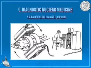
The development of large area scintillator crystals led to the development of the Gamma Camera which allowed to scan over larger areas with high spatial resolution. The Gamma camera consists out of a large area NaI crystal with a lead collimator which allows only transmission of radiation from a particular point. The detector device can be moved (rectilinear or linear) to cover larger areas. To obtain a reliable image the scanning speed vscan[cm/s] must be limited that the count rate per position Ip[counts/s] (in the photopeak) is high enough to allow sufficient information density ID [counts/cm2] in the recorded image for each scan (with a line space [cm]).
The figure shows an image from a rectilinear scan showing cancer in the upper left lung using a 67Ga citrate. The cancer is indicated by a high -countrate Tpwhich is translated into a high density of counts ID.
The thickness of the lead collimator, the orientation and size of the holes as well as the distance between source and collimator define the resolution and sensitivity of the camera. Decrease in z and d improves the spatial resolution, while the length of the collimator holes is less influential (typically determined by the thickness of the lead necessary to absorb the -radiation). The geometrical efficiency €gof the parallel hole collimator is given by the length of the holes L, the hole diameter d, the thickness of the lead between the holes l, and a constant K which depends on the shape of the holes (e.g. K=0.26 for hexagonal holes): For a parallel hole collimator the spatial resolution Rcis determined by the length of the holes L, the hole diameter d, and the source to collimator distance z:
The geometrical efficiency does not depend on the source-collimator distance z. However, if z >> L and d >> t there is a direct correlation between geometrical efficiency and resolution:
The figure shows system resolution and system efficiency for different collimator systems: LEAP low energy parallel hole collimator LEHR low energy high resolution parallel hole collimator Fan Beam diverging multi hole collimator Pin Hole single hole collimator LEUHR low energy ultrahigh resolution collimator
The tables give typical examples for parallel hole collimator types and the corresponding resolution and sensitivity.
Detectors for the Gamma-Camera are in most cases large area scintillator detectors, typically Nal-crystals with up to 50 cm diameter and 6-12 mm thickness which emit a blue green light of =415 nm. A typical spectrum for -radiation of E 150 keV indicates a resolution of 10-12 %. Phototubes are closely packed and optically coupled to the scintillator crystal to achieve high light collection efficiency. The typical arrangement gives an hexagonal array for 7, 19, 37, up to 61phototubes.
To process the data only the photopeak information is necessary. Information about scattered -rays gives unnecessary background intensity. A single channel analyzer which filters out only signals of the right energy is therefore used to clean the data and create a logic signal (TTL-pulse).
To obtain a two-dimensional image the data are recorded as a function of the x-y position of the signal. This requires a resistor network to obtain positional information for the intensity distribution. The x,y data is then digitized using ADCs and form a 2D-matrix which represents the image. The x,y data is gated by the logic signal to remove background events originated by scattered -rays from the event matrix.
As example for the spatial distribution the figure shows a image of a uniform radionuclide distribution, obtained with a fully operating gamma camera and (at the right hand side) an image obtained with a gamma camera with one defect photo tube. Uniformity images are necessary to test the quality of the equipment. As examples are shown two images of the brain, based on the blood distribution in the brain. Left the brain of a patient suffering from stroke. The arrow indicates the lack of cerebral blood volume. Right a patient suffering from brain tumor.
Saha textbook………………. Chapter 3 Dr. Funk Skip chapter 4 Statistics Chapter 5 Production of Radionuclides Chapter 6 Interaction of Radiation with Matter Chapter 7 Gas detectors Chapter 8 Scintillation and Semi-conductor detectors