Download
1 / 23
240 likes | 426 Views
DNA & RNA Isolation. DNA very stable and long; easy to inactivate deoxyribonucleases RNA more subject to degradation by RNases General steps: Isolate cells, Lyse cells, Remove proteins, Isolate pure nucleic acid

E N D
DNA & RNA Isolation DNA very stable and long; easy to inactivate deoxyribonucleases RNA more subject to degradation by RNases General steps: Isolate cells, Lyse cells, Remove proteins, Isolate pure nucleic acid DNA - during isolation exclude free Mg2+ (cofactor for DNases and can be excluded by adding 10 mM EDTA as a chelator and by use of SDS) and avoid rough treatment (rigorous pipetting, vortexing should be avoided if DNA > 10,000 bp) 1. Isolate cells by centrifugation 2. Lyse cells by adding SDS for euks or lysozyme + SDS for proks also French press and sonication 3. Remove protein by adding protease (proteinase K) and extracting with phenol (denatures proteases and other proteins); Nucleic acids are precipitated by adding NaOAc and ethanol (spool genomic DNA or centrifuge smaller DNA) 4. Isolate pure DNA by equilibrium density gradient ultracentrifugation which uses the fact that DNA has a characteristic density (1.7 g/mL); density gradient of CsCl, ultracentrifuge, ethidium bromide added RNA - need to get rid of RNases with phenol extraction and presence of divalent metal ions is helpful • Hard to isolate RNA from tissues b/c does not homogenize so can use guanidinium isothiocyanate (dissolves tissue) • Separate tRNA from rRNA b/c different solubilities, rRNA ppt’d and tRNA purified by anion exchange chromatography • purify mRNA with poly A tail by affinity chromatograpy (oligo-dT cellulose chrom)
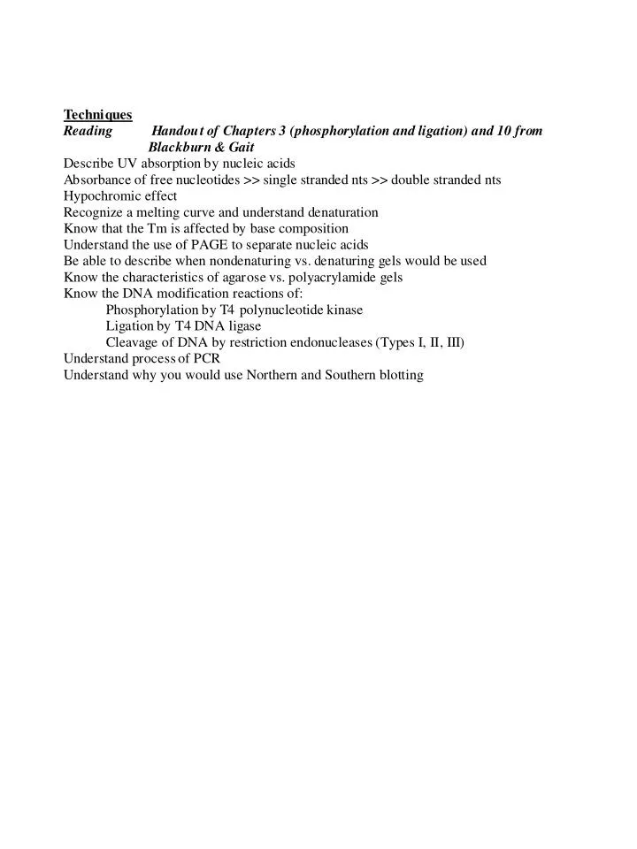
UV spectroscopy DNA and RNA nucleotides have strong UV absorption at lmax = 260 nm Absorption due to complex electronic transitions in Pu/Py rings Free nucleotides have highest absorption Single stranded nucleic acids have medium absorption Double stranded nucleic acids have low absorption Hypo- and hyperchromicity Melting temperature (Tm) depends on base composition, [salt] and counterions
Nucleotides & Nucleic Acids Chemistry - denaturation High temp/pH No covalent bonds broken Normal temp/neutral pH Partially denatured (12 bp) QUICK SLOW
Nucleotides & Nucleic Acids Chemistry - denaturation Lowest absorption of UV light HYPOCHROMIC EFFECT (can monitor transition from ds to ss) Lower absorption of UV light than free nucleotides
Nucleotides & Nucleic Acids Chemistry - heat denaturation related to absorption
Nucleotides & Nucleic Acids Chemistry - denaturation Electron microscopy
Fluorescence Emission of radiation given out as molecule returns to its ground state from an excited electronic state Can add fluorophore to DNA/RNA and use FRET FRET Fluorescence Resonance Energy Transfer energy passed over a distance donor molecule is a fluorophore - excite just it and it transfers the energy of an absorbed photon to an acceptor molecule can look at topology
Structure Determination CD - circular dichroism IR - infrared spectroscopy NMR - nuclear magnetic resonance spectroscopy X-ray crystallography NAIM - nucleotide analog interference suppression
Separation Centrifugation Separate by differences in size, viscosity or density Equilibrium centrifugation (buoyant density) - use CsCl Differential - separates by differences in size Sucrose density gradient DIFFERENTIAL CENTRIFUGATION ISOPYCNIC (SUCROSE-DENSITY) CENTRIFUGATION Tissue homogenization centrifugation Low speed Supe to medium speed Supe to high speed Tissue homogenate Sample Supe to very high speed Sucrose gradient Pellet of whole cells, nuclei, cytoskeleton, plasma membrane Less dense Fractionation More dense Pellet of mitochondria, lysosomes, peroxisomes Supernatant contains soluble proteins Pellet of micorsomes (fragments of ER), small vesicles Pellet of ribosomes, large macromolecules
Separation Electrophoresis DNA/RNA molecules move under the influence of electric field Migration based on size, charge, shape (nondenaturing vs. denaturing)
Protein Purification and Analysis Electrophoresis Use gels made of crosslinked polymer polyacrylamide Proteins/DNA/RNA migrate based on their charge-to-mass ratio (denaturing) Proteins/DNA/RNA migrate based on their charge-to-mass ratio and their shape (nondenaturing) Purification of RNA polymerase Steps 1 2 3 4 5 6
Protein Purification and Analysis Denaturing Gel Electrophoresis Used to estimate purity and molecular weight Denature protein or nucleic acid by adding SDS/formamide/urea (separate by size only) Electrophoresis of denaturing polyacrylamide gel Visualize DNA/RNA by UV shadowing 20% denaturing acrylamide Xylene Cyanol (28-mer) Bromphenol blue (8-mer)
Separation Electrophoresis Agarose PAGE (polyacrylamide) Separate large mcs (2000 kbp) separate smaller mcs (< 1 kbp) Solidify crosslink Nondenaturing denaturing vs, nondenaturing Ethidium bromide EtBr, radioactivity, fluores Mobility observed based on migration of dyes Agarose gel - can detect difference between supercoiled, relaxed and linear DNA PAGE - can detect bent DNA since it migrates slower than straight
DNA shape Pulsed field electrophoresis efficient resolution of very large DNA (> 1000 kbp) alternating polarity field + pulse longer Modeling 3D visualization of structure Microscopy look at structure of large systems
Separation Chromatography 1. Column is packed with material (resin) that can absorb molecules based on some property (charge, size, binding affinity, etc.) 2. Molecules washed through the column with buffer 3. Fractions are taken, at some point your molecule will elute 4. May have to change buffer to get elute tightly bound molecules
DNA/RNA modification 5’-phosphorylation enzyme T4 polynucleotide kinase transfers g-phosphate of ATP to 5’-OH terminus of DNA/RNA (ss or ds) T4 polynucleotide kinase Phosphatase hydrolyzes monoesters to produce Pi and alcohol most are nonspecific alkaline phosphatases found in bacteria, fungi, and higher animals (not plants) and remove phosphates from polynucleotides, carbs, phospholipids catalytic sequence Asp-Ser-Ala (serine proteases)
DNA/RNA modification Ligases catalyze formation of a phosphodiester linkage between 2 chains Need 5’-phosphate and 3’-OH and in reaction get PPi hydrolysis
DNA/RNA modification Restriction endonucleases Type I - trinucleotide and tetranucleotide sequence separated by 6 nt, cleavage < 7000 bp away Type II - palindromic, cleavage site within or real close Type III - asymmetric pentanucleotide sequence, cleavage, < 25 bp
DNA/RNA modification Nucleases
PCR After cycle 30, > 1 billion identical molecules (230 = 1.07 x 10⁹)
Northern and Southern Blotting Detection of specific nucleic acid sequences Process: 1. if genomic DNA - cut into workable pieces with restriction endonucleases 2. Separate by electrophoresis 3. Blot onto nitrocellulose 4. Gene of interest detected on filter by hybridizing a complementary nucleic acid strand labeled either with radioactivity or an affinity label such as biotin Southern - transfer DNA onto filter Northern - transfer RNA onto filter (have to keep denatured so use formaldehyde) In situ hybridization - cells and organisms smaller than 1 mm are fixed using formaldehyde; larger organisms are sliced into thin sections Specimens are probed with radioactive nucleic acid