Download
1 / 43
480 likes | 1.1k Views
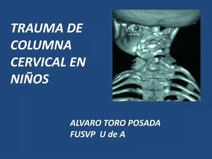
TRAUMA DE COLUMNA CERVICAL EN NIÑOS. ALVARO TORO POSADA FUSVP U de A. HISTORIA CLÍNICA TRAUMA CERVICAL INFANTIL. Torticolis Limitada movilidad Espasmo nucal R eflejos anormales C lonus R igidez. Hipotensiòn < frecuencia cardíaca Respiración diafragmática P riapismo

E N D
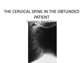
TRAUMA DE COLUMNA CERVICAL EN NIÑOS ALVARO TORO POSADA FUSVP U de A
HISTORIA CLÍNICA TRAUMA CERVICAL INFANTIL • Torticolis • Limitadamovilidad • Espasmonucal • Reflejosanormales • Clonus • Rigidez • Hipotensiòn • <frecuenciacardíaca • Respiracióndiafragmática • Priapismo • Alteracióntemperatura
JUSTIFICACION • Porque diferente del adulto? • Como lo evalúo? • RX, TAC o RM y cuando? • Manejo inicial • Excepciones
TRAMPAS EN EL TRAUMA CERVICAL INFANTILHaizlipJA; ScherrerPD • “no creoquenecesite un collar cervical, estácaminandopordonde se accidentó” • “hay quetransportarlo en unacamillarígidacomo un adulto, con correas, estásegurosiestáquieto“ • “ tiene 5 años, como no le duele el cuello, piensoque no necesitainmovilización con collar”
TRAMPAS DEL TRAUMA CERVICAL • “estoycasiseguroque la líneaqueveo en los RX esunaplaca de crecimiento, puesespequeña y lasfracturas a estaedad son muyraras” • “el técnico de RX no podrátomar en esteniñounasplacaspues no se quedaquieto, menosunabocaabiertaparaodontoides” • “el niñoenviadoparaplacas en flexión y extensióntiene dolor, lashago?”
TRAMPAS TRAUMA CERVICAL • “para mayor seguridadtodoniño con trauma cervical deborealizaruna TAC” • “ todaslasradiografías son normales y ellapareceestarbien, puedodecirle a los padres que no se preocupen de nada” • “como el niñoqueestáinconcientetiene RX y TAC cervical normal, puedodejarlo sin collar”
Trauma de columna cervical Bollini • HC por el paciente o acompañantes • Mecanismo • Signos y síntomas Nx • Intoxicación o medicamentos • Antecedentes de trauma, enfermedades o síndromes • 35-45% de Tx de columna • 0.34% de traumas infantiles • Cervicalgia • Torticolis fija • Dolor • Deficit neurológico
Trauma cervical 65% H 35 %F • RX AP, lat, bocaabierta, flexiònextensiòn ? • TAC si hay imagen de fractura • RM • Acctto 48% • Deportes 35% futbol, clavados, fútbol americano, lucha libre • Caídas 15% • 52% cervical superior • 40% TEC y 60% C0C1C2 • 30% Cx Pediatric cervical spine injuries: report of 102 cases and reviewof theliteratureMOHAMMED A. ELERAKY, M.D., NICHOLAS THEODORE, Neurosurg (Spine 1) 92:12–17, 2000
Errores DX <8a 24% • ≥9 a 15%. • > uniónC0C2 • Predisponen: • no familiarizado con patología, • anatomía, • variantes de lo normal The misdiagnosis of acute cervical spineinjuries and fractures in infants and children:the 12-year experience of a level I pediatricand adult trauma center, Anthony M., Childs NervSyst (2005) 21: 122–127 Niño 2ª Sciwora
La incidencia de SCIWORA en niñosva de 4% a 67% • Predisponentes : anomalias , ciertaspatologìas Down, KlippelFeil, Chiari, neoplasias, infecciones, ttnometabòlico • El fulcroestá C5C6 en adolescentes, C2C3 en niñosmenores Cervical Spine Injuries in Children: A Review of 103 PatientsTreated Consecutively at a Level 1 Pediatric Trauma Center, Rebeccah L. Brown of Pediatric Surgery, Vol 36, No 8 (August), 2001:
A Prospective Multicenter Study of Cervical Spine Injury in ChildrenPeter Viccellio, MD*; Harold Simon, MD‡; Barry D. Pressman, MD§; Manish N. Shah, for the NEXUS Group PEDIATRICS Vol. 108 No. 2 August 2001 • Diferenciasanatòmicas entre niños y adultoshasta los 8a y un pocohasta los 12a– • Nexus estudioprospectivoobservacional • >% niñosgrandescaracterìsticascomoadultos • SCIWORA esmuyraro en niñoscentromedular, Brown Sequard, sìndromemedular posterior • Maltratoinfantil
POSNA 2012 • Orientaciòn de las facetas màs horizontal • Fusiòn de los centros de osificaciòn de la odontoides entre 3-6ª • A menor edad mayor movilidad de la columna que la mèdula • Hipermovilidad y > tamaño de la cabeza, > vulnerabilidad cervical superior. • TEC es la lesiònmásfrecuenteasociada en niñospequeñoshasta 50% • RX en flexiònextensiòn cervical si hay hallazgos sospechosos • Aufdermaurlesionesplaca de crecimiento y SCIWORA y estudiosrecientes de RM muestranademáslesionesligamentarias Radiography of Cervical SpineInjury in Children:Are Flexion–ExtensionRadiographsUsefulforAcute Trauma?Jerry Raphael Dwek Christine B. Chung AJR:174, June 2000
COLUMNA CERVICAL EN NIÑOS • Reconocercentros de osificación y lasfisis • La sedoluxación C2/3 común en <8a, • Siga la líneaespinolaminarSwischuk • Evaluacióntejidosblandos anterior
INDICACIONES DE TAC RM • Estado mental alterado • TAC 98% sensibilidad y especificidad (irradia x 10) • IRM SCIWORA • RM edema, lesiones ligamentarias C0C2, subluxación, distracción, contusión medular, hemorragia, protrusión discal lesiones ligamento longitudinal, fracturas ocultas
Se solicitarà una RM cuando se tenga un niño con trauma cervical, o fractura de esta zona, cuando haya disociación entre los hallazgos clìnicos y RX • MRI in the assessment of the supportive soft tissues of the cervical spine in acutetrauma in children M. D.Keiper R. A.ZimmermanNeuroradiology (1998) 40: 359±363 4a, TEC, trauma cervical C0C1C2 distracción sin lesión ósea. RM Sciwora
MANEJO DEL TRAUMA CERVICAL • ABC • Proteger la columna cervical, todoniño con trauma cervical o de cràneo, politraumatizado o con lesiónneurológica, debe ser tratadocomosituviera un trauma de columna cervical Objetivos: Estabilizarlaslesionesprimarias y prevenirlaslesionessecundarias • entre 3% - 25% de lesiones se presentandurante el transporte o manejoinicial • El tratamientodefinitivo no es un objetivoinicial
TRATAMIENTO DEL TRAUMA CERVICAL • Estabilizar la lesiónprimaria y prevenirlassecundarias • No hay unaguía universal • Evaluación con neurocirugía • Reduccióncerrada y halo • Cirugía en algunoscasos, ligamentos • Esteroides no hay guías en los niños
INMOVILIZACIÓN DE LA COLUMNA CERVICAL PARA EL TRANSPORTE DE NIÑOS • La cabeza con mayor tamañoincrementa la flexión en unatablacomún • Levantar los hombros o planodeprimido en la cabezacolumna en neutro
INMOVILIZACIÓN DE LA COLUMNA CERVICAL • Collar duro • Evitar collar grandepueshiperextiende • Receso en la mesa de transportepara la cabeza, o realce de los hombros • Correas en la frente, el mentón, hombroscaderas, muslos y tobillos • Pendientesi se presentanvómitos
Radiographic studies obtained in a 2-year-old child. Left: Preoperative radiograph of the patient in a halo brace, demonstrating atlantooccipital dislocation. Center and Right: Postoperative flexion (center) and extension (right) radiographs obtained 6 months after occipitocervical fusion in which a Steinmann pin and bone graft were used, demonstrating an absence of pathological motion and an increase
Odontoid Process (Dens) Fracture Fracture through base of dens. Dens and C1 posterior to C2
The ABCS of Radiographic Cervical Spine Evaluation • A. Alignment:Lordotic curves, malalignment, subluxation, distraction. • B. Bones: Fractures, anterior and posterior cervical columns, ossification centers • C. Cartilage: Intervertebral disk spaces, ossification centers • S. Soft Tissues: Prevertebral, predental spaces.
Cervical Spine Injuriesin Children Arturo S. Gastañaduy M.D. Associate Professor of Pediatrics Louisiana State University Health Sciences Center July 2010
Odontoid Fractures • Better seen in open mouth views. • Type I: fracture at the tip of the odontoid. • Type II: Fracture at the base of the odontoid. • Type III: Fracture extends to the body of the odontoid
Posterior Cervical Line (PCL) of Swischuk • PCL connects the anterior aspect of the spinous processes of C1 and C3 • If subluxation of C2 on C3, draw PCL • (A) No subluxation. PCL cannot be applied • (B) Subluxation: Anterior aspect of C2 spinous process misses PCL >2 mm (hangman’s fracture) • (C) Pseudosubluxation: Anterior aspect of C2 spinous process <2 mm or touches PCL
Limitations for the routine use of the CT and MRI in the evaluation of cervical spine in children • Cervical spine injuries are rare in children • CT radiation dose is 10 times > plain films • CT is more costly • MRI availability is limited • MRI difficult for critically ill child
Anatomy – C1 • 3 ossification centers at birth – body and 2 neurocentral arches • Neurocentral synchondroses (F) fuse at about 7 years of age
Anatomy – C2 • 4 ossification centers at birth – body, 2 neural arches, dens • Neurocentral synchondroses (F) fuse at age 3-6 years • Synchondrosis between body and dens (L) fuses age 3 – 6 years • Thus no physis / synchondrosis should be visible on open mouth odontoid view in child older than 6 years
Anatomy – C2 • Summit ossification center (H) appears at age 3 – 6 and fuses around age 12 • Do not confuse with os odontoideum