Download
1 / 51
540 likes | 587 Views
Explore the essential components of the circulatory system, including the heart, blood vessels, and blood. Learn about plasma, red and white blood cells, platelets, and their functions in maintaining homeostasis. Discover the production of formed elements and the critical role of blood cells in transporting oxygen and nutrients throughout the body.

E N D
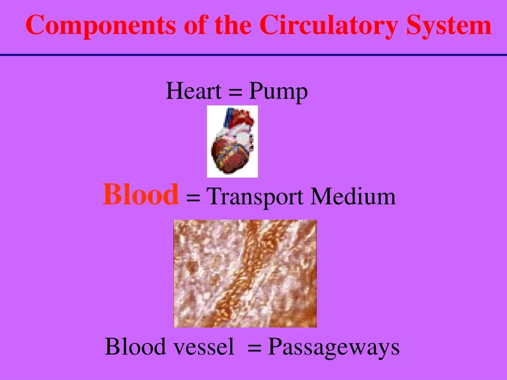
Components of the Circulatory System Heart = Pump Blood = Transport Medium Blood vessel = Passageways
Maintains Homeostasis Functions of Blood Transports: Defense: • Nutrients • Foreign organisms • Electrolytes • Injury/infection • O2 & CO2 • Clotting process • Waste Products • Hormones • Body temperature
Components of Blood Blood is a mixture of cellular components suspended in plasma: 1. Erythrocytes (RBCs) 2. Leukocytes (WBCs) 3. Thrombocytes (platelets) Total Blood Volume: 8 % of body weight 2.75 / 5.5 liters of blood is plasma (remaining is the cellular portion)
Blood vessel White blood cell Red blood cell platelet Plasma
Hematocrit “Packed Cells” • RBCs heaviest – packed at bottom after centrifugation • Average 45% for men / 42 % for women • Important clinical diagnostic marker • Anemia = Low percentage of erythrocytes • Hematocrit – mostly RBCs b/c they are the most abundant type of blood cell (99%) • Plasma = rest of blood not occupied by RBCs (55% of whole blood for males/ 58% for females)
Plasma = Less Dense Platelets / WBC’s Hematocrit “Packed Cells” More Dense Separation of Components
Components of Plasma • Blood plasma Consists of: • Water 90% • Plasma Proteins 6-8 % • Electrolytes (Na+ & Cl-) 1% • Other components: • Nutrients (e.g. Glucose and amino acids) • Hormones (e.g. Cortisol, thyroxine) • Wastes (e.g. Urea) • Blood gases (e.g. CO2, O2)
Functions of Plasma 1. Water: * Transport medium; carries heat 2. Electrolytes: * Membrane excitability * Osmotic distribution of fluid b/t ECF & ICF * Buffering of pH changes 3. Nutrients, wastes, gases, hormones: • No function – just being transported 4. Plasma Proteins (See Next Slide)
Plasma Proteins Plasma Proteins: (albumins, globulins, fibrinogen) 1. Maintaining colloid osmotic balance (albumins) 2. Buffering pH changes 3. Transport of materials through blood (such as water insoluble hormones) 4. Antibodies (e.g. gamma globulins, immunoglobulins) 5. Clotting factors (e.g. fibrinogen)
3 Cellular Elements of Blood 1. Red Blood Cells 2. White Blood Cells 3. Platelets
Production of Formed Elements • Hematopoiesis or hemopoiesis: Process of blood cell production • Stem cells: All formed elements derived from single population • Proerythroblasts: Develop into red blood cells • Myeloblasts: Develop into basophils, neutrophils, eosinophils • Lymphoblasts: Develop into lymphocytes • Monoblasts: Develop into monocytes • Megakaryoblasts: Develop into platelets
1. RBC’S (Erythrocytes) • Shape - a biconcave disc with large surface area • Can change shape • No Nucleus / organelles • Contains hemoglobin Primary Function = Transport oxygen from the lungs to the cells of the body & assist with CO2 removal
Mechanism of Transport HEMOGLOBIN * 4 Heme Molecules = * 4 Oxygen Molecules *Oxygenated Hemoglobin Bright Red (systemic) *Deoxygenated Hemoglobin Blue (venous circulation)
RBC’S (Erythrocytes) cont… • Lack intracellular organelles necessary for cellular repair, growth, division • Short Life Span (~120 days) • Aged RBC • Fragile - prone to rupture • Ruptured RBC’s are destroyed in spleen • Phagocytic WBC’s “clear the debris”
Formation of New RBC’s Ruptured cells must be replaced by new cells by a process called……… ..Erythropoiesis Secretion of the hormone erythropoietin New RBC’s (and platelets & leukocytes) are produced in the Bone Marrow
Too few, Too many • Anemia – low hematocrit (below-normal oxygen-carrying capacity of the blood) • Nutritional, pernicious, aplastic, renal, hemorrhagic, hemolytic • Polycythemia- abnormally high hematocrit (too many RBCs in circulation) • Primary, secondary
WBC’ s RBC’s
2. White Blood Cells (Leukocytes) • Mobile units of body’s defense system: • “Seek and Destroy” Functions: • Destroy invading microorganisms • Destroy abnormal cells (ie: cancer ) • Clean up cellular debris (phagocytosis) 3. Assist in injury repair
5 - Types of WBC’s Granulocytes Agranulocytes Each WBC has a specific function
Blood Cell Origin and Production Bone Marrow Circulation Figure 11-8
Types of WBC’s Polymorphonuclear Granulocytes • Neutrophils • Eosinophils • Basophils
1. NEUTROPHILS * 50-70% of all leukocytes (most abundant of WBC’s) * Important in inflammatory responses * Phagocytes that engulf bacteria and Debris
2. EOSINOPHILS * 1-4% of the WBC's * Attack parasitic worms * Important in allergic reactions
3. BASOPHILS * 0.5% of the WBC's * Release histamine and heparin * Important in Allergic Reactions * Heparin helps clear fat from blood
Types of WBC’s Mononuclear Agranulocytes 4. Monocytes 5. Lymphocytes (B and T cells)
4. MONOCYTES * 2-6 % of the WBC's * Exit blood (diapedesis) to become macrophages * Phagocytic = defend against viruses and bacteria
5. LYMPHOCYTES * 25-33 % of the WBC's * B-lymphocytes: Produce Antibodies * T-lymphocytes: Directly destroy virus- invaded cells and cancer cells
Platelets Blood vessel White blood cell Red blood cell Plasma
3. Platelets (Thrombocytes) * Cell fragments bound to megakaryocytes * “Bud Off” and are released into the blood
Function of Platelets • Stop bleeding from a damaged vessel * Hemostasis • Three Steps involved in Hemostasis 1. Vascular Spasm 2. Formation of a platelet plug 3. Blood coagulation (clotting)
Steps in Hemostasis • *DAMAGE TO BLOOD VESSEL LEADS TO: • Vascular Spasm: • Immediate constriction of blood vessel • Vessel walls pressed together – become “sticky”/adherent to each other • Minimize blood loss
Steps in Hemostasis 2. Platelet Plug formation: (figure 11-10) a. PLATELETS attach to exposed collagen b. Aggregation of platelets causes release of chemical mediators (ADP, Thromboxane A2) c. ADP attracts more platelets d. Thromboxane A2 (powerful vasoconstrictor) * promotes aggregation & more ADP • Leads to formation of platelet plug !
Figure 11-10 (+) Feedback promotes formation of platelet Plug !
Thrombin Final Step in Hemostasis • Blood Coagulation (clot formation): • Transformation of blood from liquid to solid • Clot reinforces the plug • Multiple cascade steps in clot formation • Fibrinogen (plasma protein)Fibrin “Clotting Cascade”
Factor X Thrombin in Hemostasis Figure 11-11
Clotting Cascade • Participation of 12 different clotting factors (plasma glycoproteins) • Factors are designated by a roman numeral • Cascade of proteolytic reactions • Intrinsic pathway / Extrinsic pathway • Common Pathway leading to the formation of a fibrin clot !
X Hageman factor (XII) inactive active CLOT !
Clotting Cascade • Intrinsic Pathway: • Stops bleeding within (internal) a cut vessel • Foreign Substance (ie: in contact with test tube) • Factor XII (Hageman Factor) • Extrinsic pathway: • Clots blood that has escaped into tissues • Requires tissue factors external to blood • Factor III (Tissue Thromboplastin)
Clotting Cascade • Fibrin : • Threadlike molecule-forms the meshwork of the clot • Entraps cellular elements of the blood forms CLOT • Contraction of platelets pulls the damaged vessel close together: • Fluid squeezes out as the clot contracts (Serum)
Clot dissolution • Clot is slowly dissolved by the “fibrin splitting” enzyme called Plasmin • Plasminogenis the inactive pre-cursor that is activated by Factor XII (Hageman Factor) (simultaneous to clot formation) • Plasmin gets trapped in clot and slowly dissolves it by breaking down the fibrin meshwork Figure 11-15
Clot formation:Too much or too little of a good thing… • Too much: • Inappropriate clot formation is a thrombus (free-floating clots are emboli) • An enlarging thrombus narrows and can occlude vessels • Too little: • Hemophilia- too little clotting- can lead to life-threatening hemorrhage (caused from lack of one of the clotting factors) • Thrombocyte deficiency (low platelets) can also lead to diffuse hemorrhages
Blood Grouping • Determined by antigens (agglutinogens) on surface of RBCs • Antibodies (agglutinins) can bind to RBC antigens, resulting in agglutination (clumping) or hemolysis (rupture) of RBCs • Groups • ABO and Rh
Rh Blood Group • First studied in rhesus monkeys • Types • Rh positive: Have these antigens present on surface of RBCs • Rh negative: Do not have these antigens present • Hemolytic disease of the newborn (HDN) • Mother produces anti-Rh antibodies that cross placenta and cause agglutination and hemolysis of fetal RBCs
Diagnostic Blood Tests • Type and crossmatch • Complete blood count • Red blood count • Hemoglobin measurement • Hematocrit measurement • White blood count • Differential white blood count • Clotting


