Download
1 / 21
220 likes | 410 Views
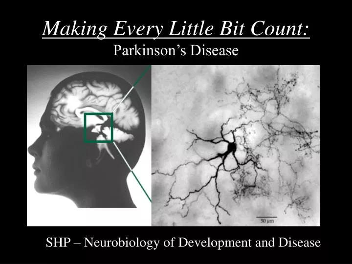
Making Every Little Bit Count: Parkinson’s Disease. SHP – Neurobiology of Development and Disease. Parkinson’s Disease. Initially described symptomatically by Dr. James Parkinson in 1817 in An Essay on the Shaking Palsy.

E N D
Making Every Little Bit Count:Parkinson’s Disease SHP – Neurobiology of Development and Disease
Parkinson’s Disease • Initially described symptomatically by Dr. James Parkinson in 1817 in An Essay on the Shaking Palsy. • Biochemical deficit and death of neurons was not characterized until the 1957 discovery by Arvid Carlsson that dopamine is a neurotransmitter. • Symptoms: • Tremor (4-7Hz) • Rigidity • Akinesia/Bradykinesia • Postural Instability (stooped posture, lack of balance) • Dementia, memory loss • Speech problems • Facial masking • PD appears to occur in approximately 2% of individuals
PD is Caused by a Loss of Midbrain Dopaminergic Neurons • Disease becomes manifest when ~80% of dopamine lost and 60% of neurons are lost. • Dopaminergic dropout can be seen in the destaining of neuromelanin in the substantia nigra • Some loss of neurons is also observed in noradrenergic (locus coeruleus), serotonergic (raphe), and cholinergic, olfactory bulb and autonomic nervous system. PD Normal http://www.path.sunysb.edu/neuropath/images/neuro_degen/neoro_degen_parkinsons_1.JPG
Dopaminergic Circuits • Mesolimbic pathway: project from the ventral tegmental area (VTA) to the cerebral cortex, nucleus accumbens, and the hippocampus. • This system appears to be involved in the dopaminergic arm of addiction. • Overactivation of dopamine in this circuit is associated with schizophrenia. • Nigrostriatal pathway: project from the substantia nigra to the striatum • This is the zone where most loss of neurons occurs in PD
PET Scan Reveals Loss of Dopamine Receptors in the Brain • Injecting patients with 18F-dopamine allows quantification of dopaminergic receptors in the brain. • PD patients have a great diminishment of receptors in the brain (as a results of DA neuron death).
MPTP • 1-methyl-4-phenyl-1,2,3,6-tetrahydropyridine • Metabolized into MPP+, which kills dopaminergic neurons in the substantia nigra by oxidative damage • Discovered in 1972 when a graduate student, Barry Kidston, incorrectly synthesized the opioid MPPP and injected the product. He began displaying classic parkinsonian symptoms within 3 days. Two years later, he committed suicide and autopsy showed characteristic loss of DA neurons in the substantia nigra. • In 1982, MPPP was manufactured illicitly in Santa Clara County, CA and distributed as a synthetic heroin. Soon after, a surge of clinical cases began to appear, some as young as 19 years old, displaying idiopathic, end-stage parkinsonian symptoms. These cases were ultimately linked to use of MPPP batches tainted with a byproduct of MPTP. • Taken up by dopamine transporters and inhibits complex-1 of the mitochondrial transport chain. MPPP MPTP
Pathways that Are Involved with PD Moore et al, 2005
Lewy Bodies • Histological analysis of PD brains reveals inclusion bodies in dopaminergic neurons, known as Lewy bodies. • These appear to be primarily constituted of alpha-synuclein, parkin, ubiquitin, and neurofilaments.
Parkin Mutation Linked to PD • The gene Parkin, originally identified in a consanguineous Pakinstani family known to have congenital early onset PD. • A mutation in parkin has been found in 50% of the cases of early onset PD. • Parkin, containing a RING finger domain, is a E3 ubiquitin ligase involved in the ligation of ubiquitin to proteins for proteosomal degradation targeting. • Dysfunction of this gene is thought to allow abnormally folded and old proteins to inappropriately linger and accumulate in the cell, perhaps leading to aggregation.
Treatment Initiatives: • Enhancement of neurotransmitter synthesis • Prevention of neurotransmitter breakdown • Replacement of lost neurons • Preventing inappropriate neural activity downstream
Levodopa (L-dopa) • A prodrug that is administered to PD patients to enhance dopamine production (since dopamine cannot cross the blood-brain-barrier) • It is transported over the blood-brain barrier by the large neutral amino acid (LNAA) transporter. • Only 5% of the drug reaches the brain due to conversion to dopamine by AAAV in the periphery. Its action here is associated with intense nausea. • AAAV inhibitors carbidopa and benzserazide inhibit peripheral production of dopamine and increases availability for CNS.
The Distribution of Levodopa Through Biological Compartments
Disadvantages of Levodopa • The half-life Levodopa is only 2hrs but ameliorates the PD symptoms for 8hrs intervals (known as the long duration effect), which is thought to be due to transmitter being stored in the DA neurons. • After 2-4yrs of treatment, patients develop a “wearing off” where the drug seems to stop working in between doses. Now the effect of the drug is dependent on serum concentration (known as the short duration effect. • Longterm use is associated with levodopa-induced dyskinesias. • Taking too much of the drug will induce a schizophrenia-like syndrome (characterized primarily by auditory and visual hallucinations).
Dopamine Agonists • Half-live of drugs are much longer than Levodopa so smoothes out drugs in the blood over time. • Directly stimulates DA receptors, circumventing issues with synthesizing neurotransmitters in dying neurons. • Some evidence that they may be neuroprotective.
Monoamine Oxidase Inhibitors (MAOIs) • MAO is the enzyme that rapidly breaks down neurotransmitters left in the synaptic cleft. • Inhibition leaves more neurotransmitter available in the synaptic cleft • Selegiline is the most prescribed because it is selective for MAOIB (inhibition of MAOIA produces “cheese effect”, where sympathicomimetics in food like tyramine can induce sympathetic overdrive).
COMT Inhibitors • When decarboxylation of levodopa by AAAD in the periphery is blocked COMT is the second route of inactivation levodopa to 3-OMD. • Inhibiting this enzyme prevents breakdown of the drug in the periphery. • Tolcapone is an example
Anticholinergics • Utilized in the treatment of PD before the discovery of dopamine (anti-muscarinics). • Only effective against resting tremors and do no improve bradykinesia and rigidity. • The mechanism of effect is not known but is thought to act on cholinergic interneurons present in the striatum.
Deep Brain Stimulation • Surgical procedure where a electrical stimulator is implanted in the chest and wires run under the skin supply current to an electrode implanted in the dysfunctional area. • For PD these electrodes are implanted in the subthalamic region. • Alleviation of symptoms are immediate on turning on the stimulator and are reversible when turned off.