Download
1 / 19
220 likes | 643 Views
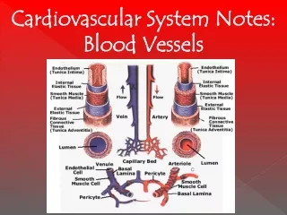
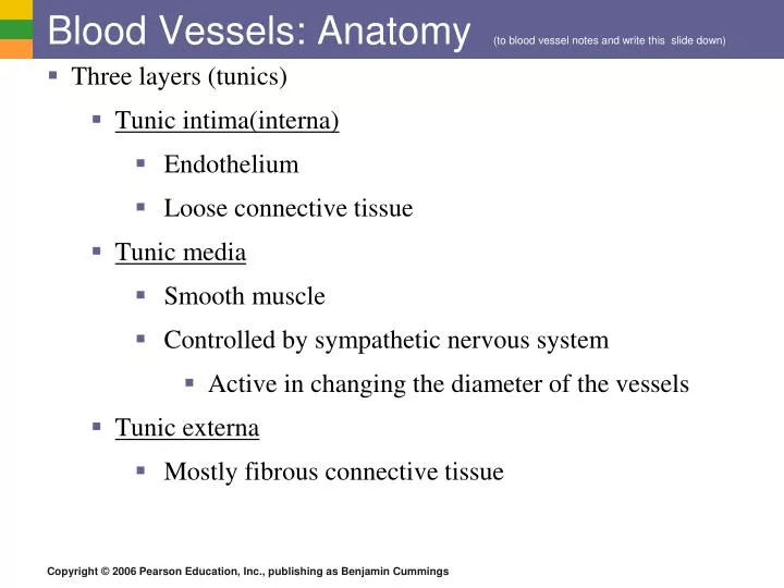
Blood Vessels: Anatomy (to blood vessel notes and write this slide down). Three layers (tunics) Tunic intima ( interna ) Endothelium Loose connective tissue Tunic media Smooth muscle Controlled by sympathetic nervous system Active in changing the diameter of the vessels

E N D
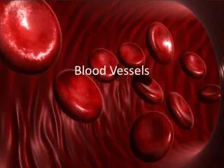
Blood Vessels: Anatomy (to blood vessel notes and write this slide down) • Three layers (tunics) • Tunic intima(interna) • Endothelium • Loose connective tissue • Tunic media • Smooth muscle • Controlled by sympathetic nervous system • Active in changing the diameter of the vessels • Tunic externa • Mostly fibrous connective tissue
The Vascular System Figure 11.8b
Variations in Blood Pressure (slide 3-6 go with blood pressure notes) Human normal range is variable Normal B.P. 140–110 mm Hg systolic 80–70 mm Hg diastolic Hypotension Low systolic (below 110) Hypertension High systolic (above 140 ) Can be dangerous if it is chronic
Blood Pressure: Factors Neural factors: Sympathetic nervous system causes vasoconstriction which raises blood pressure. Renal factors: The kidneys regulate blood volume Increased blood volume increases blood pressure Aldosterone, ADH Renin (when BP low, it stimulates aldosterone secretion)
Blood Pressure: Factors Decreased blood volume decreases blood pressure ANP(atrial natriuretic peptide) decreases blood volume. (increases urine output) Hormonal factors Epinephrine increases BP as does nicotine Poor diet choices can lead to high BP.
Blood Pressure Factors Temperature Cold-vasoconstricting effect, BP increases Cold compress to reduce swelling Warm compress: speed circulation to that area Alcohol: Decreases blood pressure, vasodilation
Capillary Beds • Capillary Beds: network of capillaries in tissues. • Microcirculation : flow of blood from arteriole to venule through the capillary bed • Oxygen and nutrients cross to cells • Carbon dioxide and metabolic waste products cross into blood Figure 11.10
Capillary Beds • Consists of 2 types vessels • Vascular shunt: vessel that directly connects the arteriole and venule • True capillaries: actual exchange vessels • Pre-capillary sphincter: surrounds root of each capillary, regulating flow of blood into the capillary • Open: blood thru true capillary • Closed: blood through shunt
Checkpoint 1. Soon after the onset of ventricular systole the: a. AV valves close b. Semilunar valves open c. First heart sound is heard d. Aortic pressure increases 2. What is the function of the fluid that fills the pericardial sac?
Capillary Exchange • Substances exchanged due to concentration gradient • Substances entering or leaving the bloodstream may take one of four routes across the plasma membrane of the single layer capillary wall • Diffusion directly through cell membranes if lipid-soluble • Enter or leave blood by endo or exocytosis if lipid-insoluble
Movement Into or Out of Bloodstream • Clefts in plasma membrane of capillaries • Limited passage of fluid and small solutes thru clefts • Fenestrated Capillaries • Pore covered by a delicate membrane • Found where filtration/absorption: priority • Kidneys/intestinal linings • Allows for free passage of small solutes and fluids
Pressure at Capillary • Blood pressure: high at arterial end • Tends to force fluids/solutes outward • Osmotic pressure: high at venous end • Draws fluid back in • Fluid: • Leaves capillary through clefts at the arterial end • Returns to blood at venous end
Diffusion at Capillary Beds Figure 11.20
Major Arteries of Systemic Circulation Figure 11.11
Major Veins of Systemic Circulation Figure 11.12
Fetal Circulation • 3 vessels • 1 umbilical vein: carries nutrient/Oxygen rich blood to fetus from placenta • 2 umbilical arteries: carries C02 and wastes from fetus to placenta • Shunt that bypasses nonfunctional liver • Ductus venosus: blood moving superiorly to the heart, by passes the liver through the ductus venosus, which connects to the inferior vena cava
Fetal Circulation • Foramen ovale: some of blood entering right atrium passes through this shunt and into the left atrium • Shunt that bypasses the immature lungs • Blood entering into right ventricle and moving into pulmonary trunk, is met by a second shunt, ductus arteriosus. • Ductus arteriosus: connects the aorta and the pulmonary trunk
Circulation to the Fetus Figure 11.15
Developmental Aspects of Cardiovascular System • Heart begins: tube-like structure • Beating/Pumping blood by 4th week of embryonic development • Atherosclerosis: Change of the arterial wall • Arteriosclerosis: results from aging • End stage of atherosclerosis • Gradual loss of elasticity in arteries • Results in hypertension, heart disease, coronary artery disease, stroke • Cardiovascular disturbances • Venous valves weaken