Download
1 / 22
220 likes | 334 Views
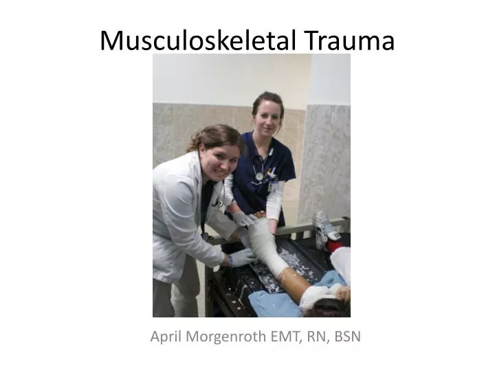
Musculoskeletal Trauma. April Morgenroth EMT, RN, BSN. Musculoskeletal System. Cartilage: softer than bone but provides structure. Bones: Provide structure and protect organs. Muscles: serve to connect musculoskeletal structures and allow movement. Tendons: connect muscles to bones.

E N D
Musculoskeletal Trauma April Morgenroth EMT, RN, BSN
Musculoskeletal System Cartilage: softer than bone but provides structure Bones: Provide structure and protect organs Muscles: serve to connect musculoskeletal structures and allow movement. Tendons: connect muscles to bones Ligaments: Support joints by attaching bones to bones us.dk.com/.../humanbody/img/image_body002.jpg
Injuries to the Joint Luxation: Total dislocation of joint. Clinical Presentation Pain Swelling http://commons.wikimedia.org/wiki/Image:Luxation_acromioclaviculaire.jpeg Decreased Range of Motion Deformity Subluxation: Partial dislocation of a joint. Possible Altered Sensation Splint dislocations in the position found, treat pain, consider anti inflammitory
Sprains and Strains • Sprain: Overstretching of the ligaments Pain Swelling Bruising 12.31.13.9/.../media/medical/hw/n5551877.jpg www.horseholistics.com/images/Img29.gif Strain: stretching or tearing of the muscle or tendon Weakness
Initial Management Rest Ice Ibuprofen or other non steroidal anti inflammatory Compression Elevate
Fractures: Simple vs. Compound Simple Fracture: The bone is fractured but the skin remains intact. http://www.nlm.nih.gov/medlineplus/ency/imagepages/8856.htm Compound Fracture: The bone is fractured and has pierced the skin. There is communication between the bone and the outside environment. http://images.medicinenet.com/images/illustrations/typical_fractures.jpg
Fractures: Non-displaced vs. Displaced • Non displaced fracture: fracture is present but the bone ends are still aligned • Displaced fracture: fracture is present but the bone ends are displaced. The fracture will need to be reduced before casting in order to heal properly.
Fracture Assessment Mechanism of Injury Swelling Crepitus Pain Deformity (may or may not be present) http://www.humanillnesses.com/original/images/hdc_0001_0001_0_img0046.jpg Decreased Range of Motion X-Ray Decreased function Always circulatory, motor, and sensory distal to the injury
Splinting • Why do it? Prevent further injury: Further movement of bone ends may cause shearing injury to surrounding tissue. Control bleeding: immobilizing the injury will help damaged blood vessels form clots to stop the bleeding. Minimize pain: moving the injured part may cause more pain and further injury.
Splinting • Remove restrictive clothing (shoes) • Immobilize the joint above and below the injury • Apply the splint with as little movement as possible to the injured part • Always check circulatory, motor, and sensory after applying a splint. • Reassess frequently. • Secure the splint snugly, enough to restrict movement but not too tight.
Compound Fractures Evaluate and treat the patient for signs and symptoms of shock. Never attempt to realign a compound fracture! Provide pain control as ordered. Gently cover the open wound with sterile dressings. This patient may need surgery to repair the fracture. Splint in the position found. High risk for infection, consider antibiotics
Rib Fractures Assessment: Pain, swelling, and/or bruising over the injured area. May or may not be deformity Pain will be worse when deep breathing, coughing, or palpated. Note, work of breathing, respiratory rate, oxygenation, heart rate, symmetry in chest rise. Flail chest: Paradoxical chest wall movement during breathing. Caused by multiple rib fractures resulting in floating rib segments. This is an emergency! www.netterimages.com/.../000/000/198-150x150.jpg
Treatment of Rib Fractures • Treating pain will help make breathing easier for the patient. • Do not place anything completely around the chest, this may restrict breathing. Have patient hug a pillow when coughing or deep breathing. • You may use the patients own arm to splint a rib fracture by sling and swath method. • Encourage coughing and deep breathing in spite of pain as this will help to prevent fluid accumulation in the lungs.
Hip Fracture In the case of Hip fracture: • Mechanism of injury • Pain, swelling, bruising, deformity • Lateral rotation or shortening of the injured leg • Check sensory, motor, circulation in the affected leg • Treat pain • Monitor vitals • Keep the patient in position of comfort • Pt may need surgery to repair the fracture www8.georgetown.edu/dml/facs/graphics/gallery.htm
Fractures of the Pelvis • Can cause life threatening bleeding • May be stable or unstable • Check stability of the pelvic girdle • Evaluate and treat the patient for shock • May have bleeding into the abdomen, check for signs and symptoms. • X-ray of pelvis and chest • An unstable pelvic fracture will need splinting. • Pt will may need surgery Picture of blanket splint
Complications Bleeding: • Bones bleed when broken • Trauma to the surrounding area from bone ends or fragments. • Check for bleeding and signs of circulation • Control bleeding as able • Evaluate and treat signs and symptoms of shock
Complications Infection • Compound fractures are at high risk for infection, treat with antibiotics prophylactically • Pain related to rib fractures may cause a patient’s breathing to become shallow and put them at risk for pulmonary complications
Complications Deep Vein Thrombosis • Evaluate circulation frequently • Pulses, capillary refill, skin temperature and color • DVT signs and symptoms: edema, redness, cyanosis, absent pulses, warm, pain • Prevention: early ambulation, physical therapy www.clotcare.com/clotcare/images/dvt4.jpg
Complications Compartment Syndrome • Increased pressure in an enclosed compartment restricts circulation and causes tissue damage and/or necrosis • Pressure may be caused by swelling and inflammation, bleeding into the space content.answers.com/.../300px-Fasciotomy_leg.jpg • Look for: signs of decreased circulation. pain, pallor, pulselessness, paralysis, pressure, and numbness • Notify MD immediately, pt may need fasciotomy, loosen splint, do not ice or elevate
Complications Fat Embolism • Occurs when fatty tissues enter the blood stream and are lodged in the narrowing blood vessels • Signs and symptoms will be similar to DVT unless the embolus is lodged in the lung or brain • If embolus is lodged in the brain, neurological deficits may result from hypoxic brain injury
Pulmonary Embolism • Obstruction of the pulmonary artery or one of its branches • Clot usually forms in the veins and then lodges in the pulmonary artery • Shortness of breath, hypoxia, tachycardia, cyanosis, anxiety, sudden death • Do: x-ray and EKG to rule out other causes, monitor vitals and oxygen saturation, provide supplemental oxygen, keep the patient calm and quiet http://jaapa.com/issues/j20060701/screen/pulmonary0706art.gif
Pneumonia • Pain related to rib fractures may cause a patient to breathe shallow and put them at risk for a atelectasis and fluid build up in the lungs which may cause pneumonia • Prolonged immobility may also lead to build up of fluid in the lungs • Prevention: coughing and deep breathing exercises, treat pain, early mobility