Download
1 / 14
200 likes | 593 Views
Microanalysis in Science and Engineering - X-ray Microanalysis. A Workshop for Middle and High School Teachers sponsored by Tennessee Technological University Center for Manufacturing Research Departments of Chemical, Mechanical, Earth Sciences and Curriculum and Instruction

E N D
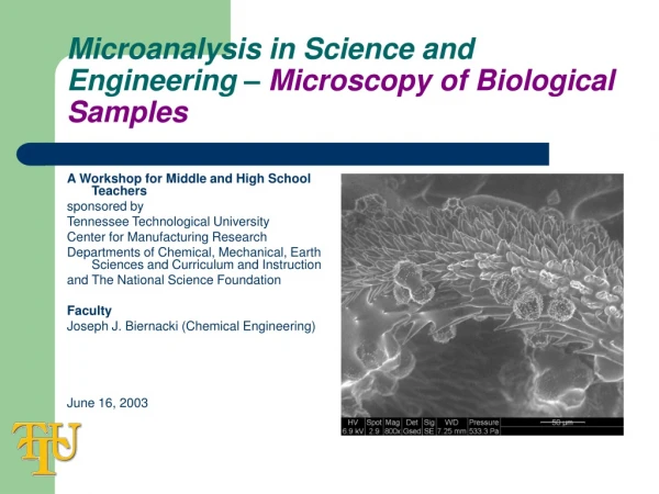
Microanalysis in Science and Engineering - X-ray Microanalysis A Workshop for Middle and High School Teachers sponsored by Tennessee Technological University Center for Manufacturing Research Departments of Chemical, Mechanical, Earth Sciences and Curriculum and Instruction and The National Science Foundation Faculty Joseph J. Biernacki (Chemical Engineering) June 16, 2003
What will we learn • What is X-ray microanalysis? • What interactions of electrons and matter are used? • How are the electron/matter interactions used to generate images and compositional information? • What linkages can be made between the “technology fundamentals” and the middle/high school science curriculum?
What is X-ray microanalysis? • X-ray microanalysis is the characterization of X-ray emissions due to the bombardment of matter with electrons. A bit of history: Mosely (1913) – the frequency of emitted characteristic X-ray radiation is a function of the atomic number of the emitting element (X-ray spectrochemical analysis) Hillier (1947) and Marton (1941) – patented an optical microscopy/X-ray spectrochemical analyzer Castaing and Guinier (1949) – the first elctron microprobe CAMECA (1956) – first commercial electron microprobe (utilized wavelength dispersive spectroscopy technology)
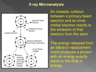
Other forms of microanalysis • When e- interact with matter, a wide range of emissions are produced including: • Elastic scattering emissions (backscattered electrons) • Inelastic scattering emissions • Secondary electrons • Continuum X-rays • Characteristic X-rays • Auger electrons • UV, IR and visible light (photons)
Suggested Curriculum Links Chemistry and Physics: quantum theory, Bohr atom incident e- ejected e- X-ray N N N scattered e- How are characteristic X-rays produces? • There are many forms of interactions, however, the production of characteristic X-rays is among the most widely used for analytical analysis.
Suggested Curriculum Links Chemistry: electron orbitals, Aufbau principle X-ray N Review of electron orbital structure • Electrons are organized into orbitals which are filled in increasing order of energy. No. of e- K 1s2 2 L 2s2p6 8 M 3s2p6d10 18 N 4s2p6d10f14 32 When an electron from a higher energy orbital makes a transition to a lower energy orbital, an X-ray is emitted with a characteristic energy. This energy is unique to the transition and element.
Suggested Curriculum Links Chemistry: quantized energy states K L M N ∞ Ka Kb K excitation Ma Mb Transitions and nomenclature • Transitions made to a shell are given the name of the shell. Transitions from one energy level above are designated as a, from two levels above b, etc.
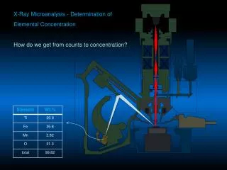
Charcteristic X-rays - examples Element Ka La Mg 1.254 Ca 3.691 .341 Fe 6.4 .705
Suggested Curriculum Links Physics: light diffraction, wave theory of matter Wavelength dispersive spectroscopy (WDS) • WDS utilizes the wave properties of X-rays to discern emitted X-ray energies. X-rays, like all wave energy, can be diffracted. Some necessary background: Diffraction of X-rays obeys Braggs law: n=1, 2, 3, … , l=X-ray wavelength, d=the spacing between atomic planes in a crystal, q=the diffraction angle incident X-rays in phase diffracted X-rays out of phase q d
incident e- X-ray detector diffracted X-rays diffraction crystal q emitted X-rays sample How does WDS work? • In WDS the emitted X-rays are diffracted by a crystal. The diffracted X-rays are counted by a detector. The intensity of the diffracted X-rays is recorded as a function of the diffraction angle.
Suggested Curriculum Links Physics: semiconductor, electron-hole pairs incident e- solid state X-ray detector emitted X-rays sample Energy dispersive spectroscopy(EDS) • EDS uses a solid state X-ray detector (Li-drifted Si crystal). Incident X-rays produce a voltage pulse that is proportional in size to the incoming X-ray energy. In this way, information about all energies (wavelengths) is gathered simultaneously producing the entire X-ray spectrum without “scanning.”
So what can we do with these technologies? • Composition point analysis.
Summary • EDS and WDS are among the most commonly used forms of SEM microanalysis. • Atoms ionized by incident e- radiation, emit X-rays upon relaxation. These X-rays are unique (characteristic) to the emitting element. • Wavelength dispersive spectroscopy (WDS) utilizes diffraction to discern between X-rays of differing wavelength (energy). • Energy dispersive spectroscopy (EDS) uses a solid state detector to convert X-rays of different energy into voltage pulses proportional to the incident energy. • WDS is more resolved than EDS, but data acquisition can be slower and the instrumentation is more expensive. • Both WDS and EDS can be used to do point analysis, composition mapping and other forms of microchemical imaging.