Download
1 / 39
390 likes | 502 Views
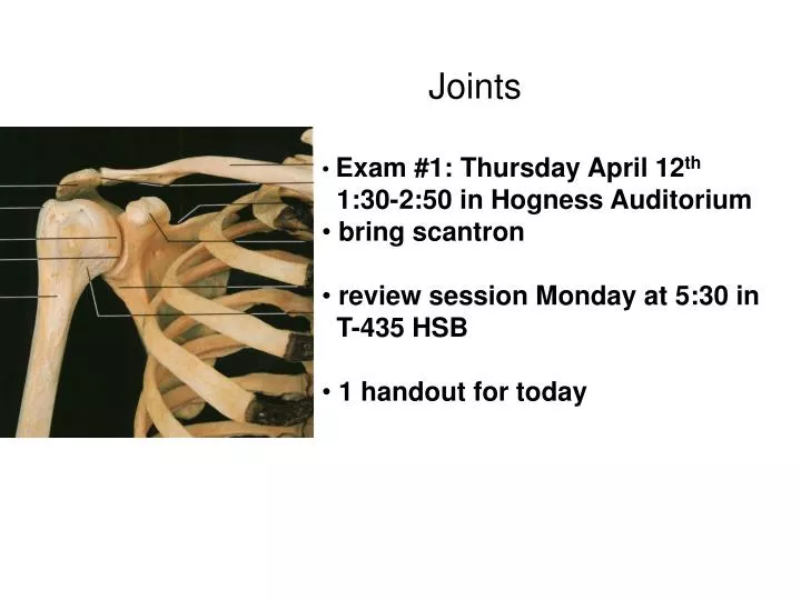
Joints. Exam #1: Thursday April 12 th 1:30-2:50 in Hogness Auditorium bring scantron review session Monday at 5:30 in T-435 HSB 1 handout for today. Name this cell. List one of it’s functions. Name this space. What is in this space?. John broke his ulna, does he need crutches?

E N D
Joints • Exam #1: Thursday April 12th • 1:30-2:50 in Hogness Auditorium • bring scantron • review session Monday at 5:30 in • T-435 HSB • 1 handout for today
Name this cell. List one of it’s functions. Name this space. What is in this space? John broke his ulna, does he need crutches? The mandible articulates with the ___________ bone.
Joints • Different type of joints • fibrous • cartilaginous • synovial • anatomy • stabilizers • temporomandibular joint • anatomy • TMJ syndrome • shoulder joint • anatomy • common injuries • knee joint • anatomy • common injuries
Joint classification • manner or type of material by which bones articulate • relates to movement Fibrous or cartilaginous synovial
Fibrous • bound by collagen • fibers • little or no movement • Cartilaginous • linked by cartilage • some movement
Cartilaginous Joints • linked by cartilage • hyaline or fibrocartilage Rib 1 vs 2-12
Synovial joints • freely moveable • articulating bones covered with • hyaline cartilage • bones separated by joint cavity • joint cavity contains synovial fluid • joint is enclosed by joint capsule
Parts of a synovial joint Ligaments Articular (hyaline) cartilage Joint (articular) capsule (Synovial membrane + fibrous capsule) Joint cavity (contains synovial fluid) Synovial fluid and warming up
Bursa • sac filled with synovial fluid • between muscles or tendons • and bones • for protection and sliding • bursitis - inflammation • Tendon Sheaths • elongated bursae • wrap around a tendon • for protection and sliding • tenosynovitis • tendonitis
How do you stabilize a synovial joint? Name & how (4 ways)
Fig. 9.6(TE Art) Ball-and-socket joint (humeroscapular) Pivot joint (radioulnar) Hinge joint (humeroulnar) Gliding joint (intercarpal) Saddle joint (trapeziometacarpal) Condyloid joint (metacarpophalangeal)
Temporomandibular joint (TMJ) Mandibular fossa • Depress & elevate • Medial & lateral excursion • Protraction & retraction
Masseter Temporomandibular ligament Temporalis TMJ syndrome
Ball and Socket joints Coxal joint Humeroscapular joint
Humeroscapular joint (Glenohumeral) • articulating bones? • flex & extend • hyperextend • abduct & adduct • medial & lateral rotation • circumduction What about protraction & retraction of the scapula?
Sternoclavicular joint • Scapular anatomy • acromion • spine • coracoid process • glenoid cavity • AC joint
Acromioclavicular (AC) joint Acromion Supraspinatus Subdeltoid bursa deltoid Glenoid cavity Glenoid labrum
Subacromial bursa Coracohumeral Glenohumeral ligaments Transverse humeral Biceps tendon
Posterior view Anterior view supraspinatus infraspinatus Teres minor subscapularis Rotator cuff muscles
Supraspinatus tendon Infraspinatus tendon Glenohumeral ligament glenoid labrum Subscapularis tendon Teres minor tendon Glenohumeral ligaments Lateral view of glenoid fossa
1. What is this? Function? 2. What is this? Function? 3. What is this? Function? 4. When the head of the humerus comes out of the glenoid fossa, what is that injury called?
Tibiofemoral joint (knee) • articulating bones • tibia – femur • (patellofemoral joint) • movements- modified hinge • flex • extend • rotation when flexed • “locking” when fully • extended
Medial meniscus Lateral meniscus Knee flexed Menisci = Fibrocartilage pads
Quadriceps tendon Prepatellar bursa Joint capsule Synovial membrane Meniscus Patellar ligament
quads quads patella Prepatellar bursa patella ? Synovial Capsule
Housemaid’s knee (from a wrestling website….) aka prepatellar bursitis
Intracapsular & extracapsular ligaments of knee Lateral (fibular) collateral lig. Ant. Cruciate lig. Post. Cruciate lig. meniscus Medial (tibial) collateral ligament Posterior Anterior
hyperflexion hyperextension
Deviations of the tibia from the midline • unequal weight distribution • unequal wear & tear • abnormal patella tracking = chondromalacia
before after
Osteoarthritis (OA) Rheumatoid arthritis (RA)
Exam #1 this Thursday here in Hogness Bring a scantron Be on time! Muscle tissue will NOT be on exam #1