Download
1 / 25
250 likes | 347 Views
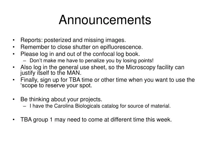
Announcements. Reports: posterized and missing images. Remember to close shutter on epifluorescence. Please log in and out of the confocal log book. Don’t make me have to penalize you by losing points!

E N D
Announcements • Reports: posterized and missing images. • Remember to close shutter on epifluorescence. • Please log in and out of the confocal log book. • Don’t make me have to penalize you by losing points! • Also log in the general use sheet, so the Microscopy facility can justify itself to the MAN. • Finally, sign up for TBA time or other time when you want to use the ‘scope to reserve your spot. • Be thinking about your projects. • I have the Carolina Biologicals catalog for source of material. • TBA group 1 may need to come at different time this week.
Immunolabeling • General problems • Immunolabeling • General considerations • Trouble-shooting • Controls for multiple antibody labeling • Filters for fluorescence • Demo and TBA
Problem 1: Bad DIC • If you don’t see a good DIC effect, first check that everything is set for DIC: • Both polarizers in • Both Wollaston prisms in • Kohler illumination set up • The knob on the second prism adjusted to neutral gray • If you still don’t have good DIC, then try this: • Pull out the prisms so you have polarization setup • Check that you have extinction (black background) • If not, then adjust bottom polarizer so that it is 90o to the top polarizer. • But the prisms back in and you should have nice DIC
Problem 2: “Posterization” of images • Java tutorial: http://micro.magnet.fsu.edu/primer/java/digitalimaging/processing/bitdepth/index.html • When describing digital images, gray-level resolution is a term that refers to the number of shades of gray utilized in preparing the image for display. Digital images having higher gray-level resolution are composed with a larger number of gray shades and are displayed at a greater bit depth than those of lower gray-level resolution. • An “over-enthusiastic” levels adjustment with Photoshop will also do this. • Oshel: Bringing down the "white" (far right) arrowhead (so perhaps also bringing up the "black" [far left] arrowhead) too much posterized the image. Looks like the bit-depth is getting truncated. • Check your images in Photoshop: resave with no levels adjustment if you see posterization.
Microtubules (Anti-tubulin) Microtubules of bovine pulmonary artery endothelial cells tagged with anti–bovine alpha-tubulin mouse monoclonal 236-10501 (A-11126) and subsequently probed with Alexa Fluor 488 goat anti–mouse IgG (H+L) antibody.
Making antibodies: Monoclonals versus polyclonals • Polyclonal antibodies bind to many sites on the antigen • Typically made in rabbit, rat or other • Monclonal antibodies bind to only one site on antigen • Always made in mice
Antibody structure 150 kD glycoprotein
ImmunolabelingProcedure • Specific antibodies used to visualize protein distribution. • Direct: specific antibody tagged with fluorochrome. • Indirect: primary (specific) antibody unlabeled, secondary antibody w/fluorochrome. • Why?
Immunolabeling References • Harlow, E. and Lane, D. (1999). Using antibodies: a laboratory manual. New York: Cold Spring Harbor Press. • Harlow, E. and Lane, D. (1988). Antibodies: a laboratory manual. New York: Cold Spring Harbor Press. • Hibbs, A.R. (2004). Confocal microscopy for biologists. Kluwer Academic. • Jackson ImmunoResearch Laboratories, Inc. www.jacksonimmuno.com
Major constraints to immunolabeling • Local antigen concentration • Large number locally • Identical antigen-binding sites • Modification of the antigen by fixation • Immobilization without change in antigen • Antibody access to the antigen • Permeability of tissue, masking of epitopes (antibody-binding site on antigen) • Antibody specificity
Permeabilizition • Allows penetration of large antibody molecules into the cellular tissue. • Typical non-ionic detergents used at 0.05-0.1% in buffers such as phosphate-buffered or Tris-buffered saline (PBS or TBS): • Triton X-100 • Tween 20 • NP-40 • Exoskeletons or other extracellular structures may require other chemical or physical disruption. • E.g. chitinous exoskeletons can be permeabilized by sonication.
Sectioned samples • Paraffin-embedded, sectioned samples • Usually not necessary for confocal • Plant tissues sometimes prepared this way for confocal • Limitation of about 200 μm for light penetration. • Cryo-sectioned samples • Sometimes the only way to preserve antigenic sites.
Methods of immunolabeling • Whole mount • Processing is done in small tubes or multi-well plates. • Adhesion of sample to slides, using poly-L-lysine or by fixation. • Spread of solutions can be limited by drawing rings with PAP pen or by using special slides. • Humidity chamber necessary to prevent drying out.
Blocking agents to prevent non-specific antibody binding • Bovine serum albumin (BSA), 0.5-2% • Skim milk, 5% • Serum (1-10%) from the same species used to raise the secondary antibody (usually goat or donkey). • Dissolved in buffer, sample treated before addition of primary antibody. • Antibody solutions usually contain blocking agent as well.
Testing specificity of a new antibody • Try antibody in immunoblotting (denatured epitope) or immunoprecipitation (native epitope) experiments to look for specific and side-reactions. • Perform appropriate controls • Negative control: confirms that a positive result in not artifactual • Preimmune or normal serum substituted for primary antibody • Secondary antibody on its own • Positive control: confirms that a negative result is not due to poor technique or reagents • Test against original target if attempting cross-reactivity • Confirm staining pattern with antibody to another epitope of the antigen. • If available, compare staining pattern in wildtype versus deletion mutation.
Variations of indirect immunolabeling Streptavidin- fluorochrome Secondary- fluorochrome Streptavidin- fluorochrome Secondary -biotin Primary- biotin Primary Primary An enzyme, e.g. horseradish peroxidase or alkaline phosphatase, can also be substituted for the fluorochrome. In this case, detection is by conversion of a substrate to a colored product.
Biotin-streptavidin BREAK: Start Staining
Immunolabeling of Drosophila embryos (Rothwell, and Sullivan, 1998. In: Drosophila Protocols, Sullivan, W. Ashburner, M. and Hawley, R.S. (eds.) Cold Spring Harbor Press, pp. 141-157) Engrailed antibody, Drosophila embryo
PBTA (1X PBS, 1% BSA, 0.05% Triton X-100, 0.02% Sodium Azide) • 10X PBS is • NaCl 80 g • KCl 2 g • Na2HPO4 14.4 g • KH2PO4 2.4 g Dissolve all components in 800 ml H2O. Adjust the pH to 7.4 with HCl. Sore at RT. • PBTA solution: Mix the following components: • 10X PBS 50 ml • BSA 5 g • Triton X-100 250 ul • Sodium azide 0.1g • Adjust volume to 500 ml with H2O.
Immunolabeling: Day 1 • Embryos have been fixed with formaldehyde and stored in methanol at -20oC. • Remove as much of the methanol as possible. • Add 500 μl PBTA solution. Allow embryos to rehydrate in this solution at room temperature for 15 minutes on a rotator. • Remove the PBTA and add 250 μl diluted primary antibody (in PBTA). Incubate on a rotator overnight at 4oC. • 1:5 engrailed, 1:5 even-skipped, 1:25 tubulin • Controls: (a) 2o antibody only, (b) neither antibody.
Immunolabeling: Day 2(In Microscopy facility) • Remove the primary antibody and rinse the embryos 3X with PBTA, allowing the embryos to settle between rinses. Wash the embryos for at least 1 hr at RT on a rotator. Longer washes and more rinses usually produce cleaner images. • Add fluorescently labeled secondary antibody (in fridg), diluted 1:250 in PBTA (250 μl total volume) and incubate 1 hr at RT on a rotator. • Remove the secondary antibody. Wash 3X with PBTA as in step 1 above. • You can cheat and wash for a total of 30 minutes. • Rinse the embryos 4X in PBS-Azide to remove the detergent. • You can cheat and do 2X washes
Mounting and Storage of Embryos • Remove as much of the PBS-Azide as possible and add 40 μl glycerol-based mounting medium (90% glycerol – 10% PBS containing 10 mg/ml N-propyl gallate to reduce photobleaching – in freezer). • Gently resuspend and transfer embryos in mounting medium to a slide using a P-200 pipetman with yellow tip cut at an angle to allow pipeting of viscous solution. • 40 μl is ideal for 22 X 22 coverslip • Place a coverslip over the embryos and seal with nail polish (sealing is optional). • Store slides flat at -20oC in the dark.
Reports (due Feb. 20) Include: • The technical information on fluorescent probe and image collection, as before. • Methods reference to Rothwell and Sullivan (1998). • Interpretation of the image, including embryonic stage, cellular and sub-cellular localization (e.g. in nuclei of dorsal epithelial cells at extended germ band stage). • On reserve: Lawrence (1992), Gilbert (2000) for staging embryos.