Download
1 / 49
740 likes | 2k Views
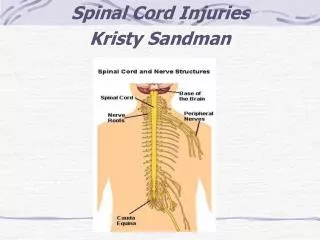
Spinal Cord Injuries. Gabriel C. Tender, MD Assistant Professor of Clinical Neurosurgery, Louisiana State University in New Orleans Staff Neurosurgeon, Touro Infirmary and West Jefferson Medical Center. Basic Anatomy and Physiology. What is the anatomy of the spinal cord on cross section?.

E N D
Spinal Cord Injuries Gabriel C. Tender, MD Assistant Professor of Clinical Neurosurgery, Louisiana State University in New Orleans Staff Neurosurgeon, Touro Infirmary and West Jefferson Medical Center
What are the clinically important ascending tracts and where do they cross over?
What are the clinically important descending tracts and where do they cross over?
At what level does the spinal cord end and why is it important?
What are the differences between UMN and LMN? (e.g., cauda equina vs. myelopathy)
Acute vs. chronic injuries;complete vs. incomplete injuries • “Acute”=sudden onset of symptoms • “Complete” ?
What is a complete spinal cord injury? • “Complete” = absence of sensory and motor function in the perianal area (S4-S5)
Terminology • Plegia = complete lesion • Paresis = some muscle strength is preserved • Tetraplegia (or quadriplegia) • Injury of the cervical spinal cord • Patient can usually still move his arms using the segments above the injury (e.g., in a C7 injury, the patient can still flex his forearms, using the C5 segment) • Paraplegia • Injury of the thoracic or lumbo-sacral cord, or cauda equina • Hemiplegia • Paralysis of one half of the body • Usually in brain injuries (e.g., stroke)
What are the important vegetative functions and when are they affected?
Reflexes • Deep Tendon Reflexes • Arm • Bicipital: C5 • Styloradial: C6 • Tricipital: C7 • Leg • Patellar: L3, some L4 • Achilles: S1 • Pathological reflexes • Babinski (UMN lesion) • Hoffman (UMN lesion at or above cervical spinal cord) • Clonus (plantar or patellar) (long standing UMN lesion)
What is and how do you determine the level of injury? • Motor level = the last level with at least 3/5 (against gravity) function • NB: this is the most important for clinical purposes • Sensory level = the last level with preserved sensation • Radiographic level = the level of fracture on plain XRays / CT scan / MRI • NB: spine level does not correspond to spinal cord level below the cervical region
Case scenario • 25 y/o white male • Fell off the roof (20 feet) • Had to be intubated at the scene by EMS • Consciousness regained shortly thereafter • Could not move arms or legs • Could close and open eyes to command • Not able to breathe by himself–totally dependent on mechanical ventilation
High cervical injuries (C3 and above) • Motor and sensory deficits involve the entire arms and legs • Dependent on mechanical ventilation for breathing (diaphragm is innervated by C3-C5 levels)
Case scenario • 19 y/o white male • Diving accident (shallow water) • No loss of consciousness • Could not understand why he could not move his legs, forearms and hands (he could shrug shoulders and elevate arms) • BP 75/40, HR 54/’ • Had difficulties breathing and required intubation a few hours after the accident
Midcervical injuries (C3-C5) • Varying degrees of diaphragm dysfunction • Usually need ventilatory assistance in the acute phase • Shock
What is the difference between spinal shock and neurogenic shock? • Spinal shock is mainly a loss of reflexes (flaccid paralysis) • Neurogenic shock is mainly hypotension and bradycardia due to loss of sympathetic tone
Neurogenic shock • Seen in cervical injuries • Due to interruption of the sympathetic input from hypothalamus to the cardiovascular centers • Hallmark: hypotension (due to vasodilation, due to loss of sympathetic tonic input) is associated with bradycardia (not tachycardia, the usual response), due to inability to convey the information to the vasomotor centers in the spinal cord
Low cervical injuries (C6-T1) • Usually able to breathe, although occasionally cord swelling can lead to temporary C3-C5 involvement (need mechanical ventilation) • The level can be determined by physical exam
So what do you expect with a cervical lesion? • Quadriplegia or quadriparesis • Bowel/bladder retention (spastic) • Various degrees of breathing difficulties • Neurogenic and/or spinal shock
Case scenario • 22 y/o Hispanic female • Motor vehicle accident (hit a pole at 60mph) • + for ETOH and THC • Short term loss of consciousness (10’) • Not able to move or feel her legs • DTRs 2+ in BUE, 0 in BLE • No bladder / bowel control or sensation • Sensory level at the umbilicus
Thoracic injuries (T2-L1) • Paraparesis or paraplegia • UMN (upper motor neuron) signs
Case scenario • 22 y/o African-American female • Motor vehicle accident • Not able to move or feel her legs below the knee • Could flex thighs against gravity • DTRs 2+ in BUE, 0 in BLE • No bladder / bowel control or sensation • Sensory level above the knee on L, below the knee on R
Cauda equina injuries (L2 or below) • Paraparesis or paraplegia • LMN (lower motor neuron) signs • Thigh flexion is almost always preserved to some degree
What is the difference between cauda equina and conus medullaris syndrome?
What is the central cord syndrome? • Cervical spinal cord involvement with arms more affected than legs • May occur with trauma, tumors, infections, etc • Traumatic lesions tend to improve in 1-2 weeks • Surgical decompression may be indicated if there is spinal stenosis
Initial Management • Immobilization • Rigid collar • Sandbags and straps • Spine board • Log-roll to turn • Prevent hypotension • Pressors: Dopamine, not Neosynephrine • Fluids to replace losses; do not overhydrate • Maintain oxygenation • O2 per nasal canula • If intubation is needed, do NOT move the neck
Management in the hospital • NGT to suction • Prevents aspiration • Decompresses the abdomen (paralytic ileus is common in the first days) • Foley • Urinary retention is common • Methylprednisolone (Solu-Medrol) • Only if started within 8 hours of injury • Exclusion criteria • Cauda equina syndrome • GSW • Pregnancy • Age <13 years • Patient on maintenance steroids
CT scan • Good in acute situations • Shows bone very well • Sagittal reconstruction is mandatory • Soft tissues (discs, spinal cord) are poorly visualized • Do NOT give contrast in trauma patients (contrast is bright, mimicking blood)
MRI • Almost never an emergency • Exception: cauda equina syndrome • Shows tumors and soft tissues (e.g., herniated discs) much better than CT scan • May be used to clear c-spine in comatose patients
Lumbar Puncture • Sedate the patient and make your life easier • Measure opening pressure with legs straight • Always get head CT prior to LP to r/o increased ICP or brain tumor
Cervical Spine Clearance • Occiput to T1 need to be cleared • ER, Neurosurgery or Orthopedics physician • If the patient • Is awake and oriented • Has no distracting injuries • Has no drugs on board • Has no neck pain • Is neurologically intact then the c-spine can be cleared clinically, without any need for XRays • CT and/or MRI is necessary if the patient is comatose or has neck pain • Subluxation >3.5mm is usually unstable
Cervical Traction • Gardner-Wells tongs • Provides temporary stability of the cervical spine • Contraindicated in unstable hyperextension injuries • Weight depends on the level (usually 5lb/level, start with 3lb/level, do not exceed 10lb/level) • Cervical collar can be removed while patient is in traction • Pin care: clean q shift with appropriate solution, then apply povidone-iodine ointment • Take XRays at regular intervals and after every move from bed
Surgical Decompression and/or Fusion • Indications • Decompression of the neural elements (spinal cord/nerves) • Stabilization of the bony elements (spine) • Timing • Emergent • Incomplete lesions with progressive neurologic deficit • Elective • Complete lesions (3-7 days post injury) • Central cord syndrome (2-3 weeks post injury)
Long term care • Rehab for maximizing motor function • Bladder/bowel training • Psychological and social support