Download
1 / 90
900 likes | 1.04k Views
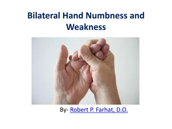
45 year old female complaint of pain and numbness of the right hand, mainly at night and during working. Carpal tunnel syndrome. Causes. Idiopathic Obesity, Oral contraceptives, Hypothyroidism, Arthritis, Diabetes,. Trauma. Lipoma , ganglion, vascular malformation. Treatment.

E N D
45 year old female complaint of pain and numbness of the right hand, mainly at night and during working
Causes • Idiopathic • Obesity, • Oral contraceptives, • Hypothyroidism, • Arthritis, • Diabetes,. • Trauma. • Lipoma , • ganglion, • vascular malformation
Treatment • Localized corticosteroid injections?????? • PT • CT Release
Triggering thumb or finger • Called stenosingtenosynovitis . Idiopathic. Or Congenital • Common disorder of later adulthood characterized by catching, snapping or locking of the involved finger flexor tendon, associated with dysfunction and pain.
Treatment • Cortisone injection • A1 poly release (surgically)
1 month old baby girl referred to you from LHC with hip click
Developmental dysplasia of the Hip (DDH) • Etiology / Epidemiology / Natural History • 1/1000 live births, left hip most common • More common in children of central European . • Etiology: multifactorial, genetic, intrauterine mechanical environment, • DDH Risk factors (five f's) • first born • female • family history • feet( breech position) • fluid(oligohydramnios )
DDH Associated AnomaliesMetatarsus adductusHyperextended knees / congenital knee dislocation Torticollis DDH Clinical Evaluation Ortolani=out-reduces Barlow=in-dislocates Asymmetric gluteal folds Galeazzi sign: apparent femoral length discrepancy when the legs are held together with the hips and knees flexed. Decreased hip abduction Ambulatory Patient: flexion contracture, gluteus medius lurch, toe walking, increased lordosisif bilateral
Treatment • Age 0-6 m=Pavlik harness • 6m-18 m= adductors tenotomy , hip Spica • >18m OR and pelvic osteotomy
Osteomyelitis • Acute, Sub acute,or Chronic. • The mechanism :exogenous or hematogeneus. • Osteomyelitis :pyogenic or non pyogenic • In infants, the infection can spread to the joint and cause arthritis. • In children, large sub periosteal abscesses can form because of the periosteumis loosely attached to the surface of the bone
Pathogenesis • leukocytes enter the infected area, and, in their attempt to engulf the infectious organisms, release enzymes that lyse the bone. • Pus spreads into the bone's blood vessels, impairing their flow, and areas of devitalized infected bone, known as sequestra, • Often, the body will try to create new bone around the area of necrosis. The resulting new bone is often called an involucrum
Cause Age groupMost common organisms • Newborns (younger than 4 mo)S. aureus, Enterobacter species, and group A and B Streptococcus species • Children (aged 4 mo to 4 y)S. aureus, group A Streptococcus species, Haemophilusinfluenzae, and Enterobacter species • Children, adolescents (aged 4 y to adult)S. aureus (80%), group A Streptococcus species, H. influenzae, and EnterobacterspeciesAdultS. aureus and occasionally Enterobacter or Streptococcus species • Sickle Cell Anemia Salmonellaspecies
Osteomyelitis • In adults, vertebrae and the pelvis. • In children, affects the adjacent ends of long bones. • Fungus :The two most common are Blastomycesdermatitidis and Coccidioideimmitis. • In osteomyelitis involving the vertebral bodies, about half the cases are due to Staphylococcus aureus, and the other half are due to tuberculosisPott's disease.
Symptoms of osteomyelitis • Pain and/or tenderness • Swelling and warmth Fever • Nausea • General discomfort, uneasiness, or ill feeling • Sinuses
Blood tests • Blood culture. • Needle aspiration • Biopsy: • Bone scan: Technetium-99 pyrophosphate
Treating and managing osteomyelitis • Drainage • Medications: • Splinting or cast immobilization • Surgery:
Duchenne muscular dystrophy (DMD) • Recessive X-linked form of muscular dystrophy, which results in muscle degeneration, difficulty walking, breathing, and death. • 1 in 3,600 -4000 boys. • Females and males are affected, though females are rarely affected and are more often carriers. • Caused by a mutation in the dystrophingene,
male children before age 5 and may be visible in early infancy. • Progressive proximal muscle weakness of the legs and pelvis associated with a loss of muscle mass is observed first. Eventually this weakness spreads to the arms, neck, and other areas. • pseudo hypertrophy (enlargement of calf and deltoid muscles), low endurance. • As the condition progresses, muscle tissue experiences wasting and is eventually replaced by fat and fibrotic tissue (fibrosis). • By age 10, braces may be required to aid in walking but most patients are wheelchair dependent by age 12.
Cardiomyopathy (DCM) , congestive heart failure or arrhythmias • A positive Gower's' sign • creatininekinase (CPK-MM) • An electromyography (EMG) shows destruction of muscle tissue Genetic testing :genetic errors in the Xp21 gene. • A muscle biopsy or genetic test (blood test) confirms the absence of dystrophin,
DNA test. DNA testing confirms the diagnosis in most cases. • Muscle biopsy A small sample of muscle tissue is extracted (usually with a scalpel instead of a needle) and a dye is applied that reveals the presence of dystrophin. Complete absence of the protein indicates the condition. • Prenatal tests
Treatment • Comprehensive multi-disciplinary care • Corticosteroids such as Prednisolone and deflazacort increase energy and strength • beta2-agonists increase muscle strength but do not modify disease progression. . • Physical therapy • Orthopedic appliances (braces and wheelchairs) • Appropriate respiratory support
Osteoarthritis • Idiopathic, primary • secondary. • Osteoarthritis causes the formation of hard, bony enlargements of the small joints of the fingers Heberden's node, • Another common bony knob (node) occurs at the middle joint of the fingers a Bouchard's node
Osteoarthritis commonly affects the hands, feet, spine, and large weight-bearing joints, such as the hips and knees
Crystal deposits in the cartilage can cause cartilage degeneration and osteoarthritis. • Uric acidcrystals cause arthritis in gout, • Calcium pyrophosphate crystals cause arthritis in pseudo gout
signs you should be aware of include: • crepitus • restricted movement or range of motion • bony enlargement • The symptoms include: • persistent knee pain • short-lived morning stiffness • functional limitation
Management • protect your joints (for example, walking is better for the joints than running). • diet and aerobic fitness exercises • Range of motion Quadriceps strengthening • Patellar taping • Lateral heel wedges • knee brace for valgus deformity • NSAID • Intra articular corticosteroids • intra-articular hyaluronic acid
Arthroscopy • Osteotomy
Definitions • Rickets : softening of bones in children due to deficiency or impaired metabolism of vitaminD, calcium or phosphorus, leading to fractures and deformity. • Predominant cause is vit. D deficiency.
Rachitic Changes Head • craniotabes(soft skull) • frontal bossing • Widening of suture • persistent fontanelae • Delayed dentition & caries
Rachitic Changes • Abdomen • Prominent • muscle weakness • Floppy baby, delayed walking • Pelvis • Narrow inlet
Rachitic Changes • Widening of wrist, knee and ankle due to physeal over growth
Rachitic Changes • Chest • Rachitic rosary • Harrison groove • Pigeon chest • Respiratory infection and atelectasis
Rickets • Vitamin D 1,25-OH helps calcium and phosphorous absorption from the intestine, increases kidney reabsorption of phosphorus, and it causes calcium and phosphorus to be released from the bone. Increasing the concentrations of calcium and phosphorus in extracellular fluid causes osteoid cells to calcify. Parathyroid hormone increases the 1-hydroxylation step of vitamin D metabolism and thereby helps regulate calcium metabolism • Ultraviolet light in the skin changes cholesterol to Vitamin D3 (i.e. cholecalciferol). Vitamin D3 then is hydroxylated in the liver to make Vitamin D 25-OH (i.e. calcidiol, the circulating reserve metabolite). It then undergoes hydroxylation again in the kidney to Vitamin D 1,25-OH (i.e. calcitriol, the active metabolite).
Management • Blood tests • Serum cacium • Serum alkaline phosphatase • Serum phosphorus • Bone x-ray • Other tests • ALP (alkaline phosphatase) isoenzyme • Calcium (ionized) • PTH • Urine calcium • Bone biopsy (rarely done)
Management • Target of therapy • Serum calcium : low - N • Serum alkaline phosphatse : high – N • Serum phosphorus : high – N
Management Treatment Vit-D deficiency state • Vit D 1,000 – 10,000 IU For 4 – 6 wks. • Vit D 300,000 IU For < 1yr of age. • Vit D 600,000 IU For > 1yr of age. • IM once OR 2 – 4 doses/day orally. All protocols followed by 400 IU/day • Calcium 1g/day.
MCQ 1-Acute osteomyelitis is commonly caused by: • a. Staph aureus. • b. S. pyogenes. • c. H. influenzae. • d. Salmonella.