Download
1 / 42
420 likes | 516 Views
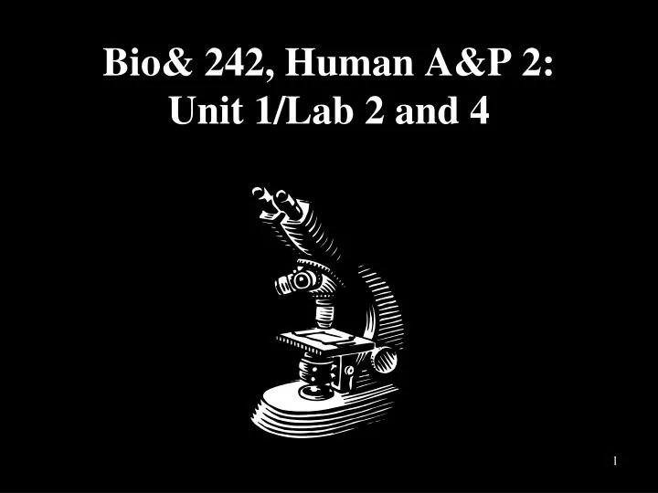
Bio& 242, Human A&P 2: Unit 1/Lab 2 and 4. Histology Slides for the GI Tract. Slides are presented in order of magnification As you view the following slides make sure you can accomplish these goals: Can you identify the organ from which the tissue sample was taken?

E N D
Histology Slides for the GI Tract • Slides are presented in order of magnification • As you view the following slides make sure you can accomplish these goals: • Can you identify the organ from which the tissue sample was taken? • Can you identify the specific structures or layers indicated by the numbered arrows or brackets? • At the end of a sequence you will find the answers to the above for each organ.
Organ? 40X 3 1 2 4 5 6 7
Organ? 100X 1 2 3 4 5
Organ ? 100X 1 4 2 3
Organ? 400X 1 2 3 4
Tissue from Esophagus slides 3-6 Slide # 3 • Nonkeratinized Stratified Squamous • Lamina Propria • General layer called the Mucosa • Muscularis Mucosae • Submucosal Mucous Gland • Submucosa • Circular smooth muscle layer • Slide # 4 • Nonkeratinized Stratified Squamous • Lamina Propria • Muscularis Mucosae • Submucosal Mucous Gland • Submucosa
Tissue from Esophagus slides 3-6 Slide # 5 • Circular smooth muscle layer • Longitudinal smooth muscle layer • Adventitia • Mucularis Externa • Slide # 6 • Nonkeratinized Stratified Squamous • Lamina Propria • Muscularis Mucosae • Submucosal Mucous Gland
Organ or Organs? 100X 1 2 1 3 4
Slides 9-10 Esophageal-Gastric Junction Slide # 9 • Junction where epithelia changes from nonkeratinized stratified squamous to simple columnar. • Simple columnar • Nonkeratinized stratified squamous • Lumen • Slide # 10 • Simple columnar lining a gastric pit • Junction where epithelia changes from nonkeratinized • stratified squamous to simple columnar
Organ? 100X 1 2 4 3 5 6
Organ? 400X 1 2 3 4 5
Organ? 400X 9 8 2 7 3 1 4 5 6
Slides 12-15 Stomach Slide # 12 Large fold is a Rugae • Gastric Mucosa • Slide # 13 • Simple columnar lining a gastric pit • Muscularis Mucosae • Lamina Propria • Oblique smooth muscle layer • Circular smooth muscle layer • Longitudinal smooth muscle layer
Slides 12-15 Stomach Slide # 14 • Muscularis Mucosae • Submucosa • Oblique smooth muscle layer • Circular smooth muscle layer • Longitudinal smooth muscle layer • Slide # 15 • Muscularis Mucosae • G-cells at the bottom of a gastric pit • Submucosa • Lamina Propria • Parietal cells in a gastric pit • Chief cells in a gastric pit • Oblique smooth muscle layer • Circular smooth muscle layer • Longitudinal smooth muscle layer
Organ? 40X 1 2 2
Organ? 400X 2 1 3
Organ? 400X 2 3 4 1
Slides 18-20 Liver Tissue Slide # 18 • Central Vein • Portal triads demonstrating the boundary of the lobule • Slide # 19 • Central Vein • Sinusoids • Hepatocyte • Slide # 20 • Branch of the Hepatic Portal Vein • Bile duct • Branch of the Hepatic Artery • Portal Triad
Organ? 40X 2 1
Organ? 100X 1 3 2
Organ? 100X 1 2
Slides 22-24 Pancreas Tissue Slide # 22 • Block of acinar cells called an acini • Pacinian Corpuscle • Slide # 23 • Block of acinar cells called an acini • Pacinian Corpuscle • Islet of Langerhans • Slide # 24 • Acinar Cell • Islet of Langerhans
Organ and folds? 40X 9 2 1 4 3 5 6 7 8
Organ? 100X 1 6 2 3 5 4
Organ? 400X 1 2 3 4 5
Slides 26-28 Small Intestine - Duodenum Slide # 26 • Villus • Lamina Propria • Lacteal • Crypt of Lieberkuhn • Submucosa • Visceral Peritoneum • Longitudinal smooth muscle layer • Circular smooth muscle layer • Plicae Circularis
Slides 26-28 Small Intestine - Duodenum Slide # 27 • Villus • Lamina Propria • Muscularis Mucosae • Brunner’s gland • Submucosa • Crypt of Lieberkuhn • Slide # 28 • Villus • Lamina Propria • Goblet cells with mucous drop • Crypt of Lieberkuhn • Muscularis Mucosae
Organ? 100X 1 2 4 6 5 3
Organ? 400X 1 2
Slides 31-32 Small Intestine - Ileum Slide # 31 • Villus • Peyer’s Patch • Muscularis Mucosae • Submucosa • Longitudinal smooth muscle layer • Circular smooth muscle layer • Slide # 32 • Muscularis Mucosae • Peyer’s Patch
Organ? 40X 1 2 3 5 4 6
Organ? 100X 1 2 3 5 4
Organ? 400X 1 3 2 4
Slides 34-36 Large Intestine Slide # 34 • Intestinal Mucosa with intestinal pits or gland • Muscularis Mucosae • Submucosa • Longitudinal smooth muscle layer • Circular smooth muscle layer • Lymph node • Slide # 35 • Intestinal pit or gland • Muscularis Mucosae • Submucosa • Longitudinal smooth muscle layer • Circular smooth muscle layer
Slides 34-36 Large Intestine Slide # 36 • Intestinal pits with goblet cells • Lamina Propria • Muscularis Mucosae • Lumen of an intestinal pit or gland
Organ or organs? 100X 3 2 1 4
Slides 39-41 Ano-rectal junction • Slide # 40 • Junction of the Rectum and Anus • Simple Columnar cells of the Rectum • Nonkeratinized stratified squamous of the Anus • Intestinal pit or gland Slide # 39 • Junction of the Rectum and Anus • Slide # 41 • Simple Columnar cells of the Rectum • Nonkeratinized stratified squamous of the Anus