Download
1 / 30
450 likes | 1.04k Views
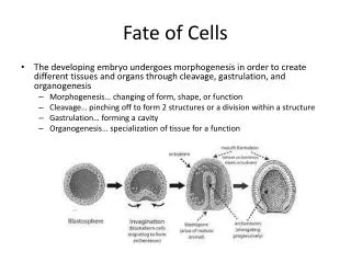
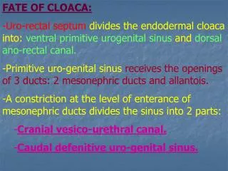
Coordination of cellular-fate processes. Dept. Physiology Chang Gung University J. K. chen, Professor. Cellular fate processes. Cells undergo various fate processes via 1. secretion of soluble signals.

E N D
Coordination of cellular-fate processes Dept. Physiology Chang Gung University J. K. chen, Professor
Cellular fate processes • Cells undergo various fate processes via 1. secretion of soluble signals. 2. secret insoluble signals that alter the physical and chemical composition of their microenvironment through modifications of the ECM. 3. they touch each other and communicate via direct cell-cell contact.
Growth factors • Growth factors may be divided into 2 groups : Cytokines– stimulate cell growth, and permit or instruct cell differentiation. Chemokines -- induce and direct cell migration.
Growth factors commonly used in cell cultures • Insulin 1922 by Banting and Best • NGF 1948 by Bueker • EGF 1962 by Cohen • IGFs 1967 by Froesch et al. • PDGF 1982 by Johnson et al. • FGFs 1987 by Gospodarowicz 1988 by Thomas • TGF-alpha 1985 by Todaro • TGF-beta 1983 by Assoian et al • VEGF 2000 by Saristo et al
The vascular endothelial cell growth factors 1. VEGF family : PlGF, VEGF-A through -D. 2. Angiopoietin family : Ang-1 through -4. 3. Ephrin family : at least one member is involved. (Ephrin-B2) Yancopoulos et al., Nature 407 (2000) JKC
VEGFs and angiopoietins, their receptor binding specificity, and some of their endothelial effects. Neuropilin (NRP-1) functions as a co-receptor for the VEGF165 isoform, P1GF-2, VEGF-B and VEGF-E. Ang-1 and Ang-4 are stimulatory ligands for Tie-2, whereas Ang-2 and Ang-3 are inhibitory Saristo et al., 2000 Oncogene Vol. 19 JKC
Lessons from gene-knockout mice SCIENCE, 277, July 1997 JKC
Hepatocyte growth factor • Stimulates division of hepatocytes, epidermal keratinocytes, renal tubular epithelial cells, and melanocytes. • Also named as scatter factor with motility and morphogenic effects. It is a paracrine factor secreted by mesenchymal cells. • It acts through cell surface tyrosine kinase receptor.
EGF(epidermal growth factor) • It stimulates the proloferation of Bone cells, smooth muscle cells, epithelial cells, heart mesenchymal cells,hepatocytes, glial cells, oral mucosal cells, fibroblasts, etc.
PDGF(platelet-derived growth factor) • A major mitogen in serum for: fibroblasts, smooth muscle cells, glial cells, chondrocytes, and other mesenchymal cells. platelet is a rich source of PDGF.
FGF(fibroblast growth factor) Present in both normal and tumor tissues. Normal tissues brain, pituitary, hypothalamus, kidney, adrenal,liver, skeletal muscle, heart, cartilage, bones, and prostate etc. Tumors hepatoma, melanoma, mammary tumor, bladder tumor,prostate tumor ,glioma ,neuroblastoma ,retinoblastoma Cells endothelial cells, smooth muscle cells, nerves, fibroblasts, macrophages.
TGF-beta • Stimulates anchorage indepent cell growth in soft agar in the presence of EGF or TGF-alpha • Stimulates matrix synthesis, and inhibits degradation. • Regulates bone remodeling by coordinated actions on chondrocytes, osteoblasts and osteoclasts • Inhibits endothelial cell proliferation, yet promotes angiogenesis in vivo.
A schematic representation of IF-gamma signal transduction pathway
PDGF and the sis oncogene • PDGFR accept signals from PDGFaa,bb, and ab. • Sis is an oncogene that encodes b chain of the PDGF
Extracellular matrix • ECM provides tissue with mechanical support • Provides cells with a substrate on which to migrate, and a place to locate signals for communication. • It is dynamic and is constantly been modified. • It has both structural and informational functions.
Malfunction in ECM signaling • Mutations in genes encoding ECM protein, ECM remodeling protein, and ECM receptors causes diseases in a variety of tissues. • Both structure and the dynamic alteration in the ECM are important in maintaining the normal tissue function.
The phosphorylation of Rb and the transcriptional activity, translocation, and degradation of P53 are regulated by the interactions of integrins and ECM components. Malfunctioning morphoregulatory control loop Rb regulates the expression of ECM-remodeling MMPs, and P53 Regulates the state of ECM through Transcriptional control of ECM components and mediators of cell-ECM signaling and ECM remodeling.
Leukocyte extravasation Activated EC express p-selectin and Paf on their surface.
Interaction between signaling mechanisms • The three signaling mechanisms are not functioning in isolation, one must be aware of potential cross-talk between signaling pathways. • Typically. All modes of communications are involved in cell and tissue processes and they thus interact or cross-talk.
相簿 由 JKCHEN