Download
1 / 23
320 likes | 1.1k Views
Female Perineum and External Genitalia. Drs. Sanaa Alshaarawy & Saeed Vohra. OBJECTIVES. At the end of the lecture, the student should be able to describe the: Boundaries of the perineum. Division of perineum into two triangles. Boundaries & Contents of anal & urogenital triangles.

E N D
Female Perineum and External Genitalia Drs. SanaaAlshaarawy & SaeedVohra
OBJECTIVES • At the end of the lecture, the student should be able to describe the: • Boundaries of the perineum. • Division of perineum into two triangles. • Boundaries & Contents of anal & urogenital triangles. • Lower part of Anal canal. • Boundaries & contents of Ischiorectal fossa. • Innervation, Blood supply and lymphatic drainage of perineum.
Perineum Perineal body • Perineum is the region of the body below the pelvic diaphragm (The outlet of the pelvis) • It is a diamond shaped area between thethighs • Boundaries: • Anteriorly Mons pubis • Laterally Medial surfaces of the thighs • Posteriorly Intergluteal folds • Contents: • Lower ends of urethra, vagina & anal canal • External genitalia • Perineal body & Anococcygeal body
Perineal Body • Perineal body is an irregular fibromuscular mass of variable size and consistency, located at midpoint of the line between the ischial tuberosities • Lies in the subcutaneous tissue, posterior to vaginal vestibule and anterior to the anal canal & anus • Forms the central point of the perineum & blends anteriorly with the perineal membrane • Function: • Gives attachment to perineal muscles • Plays an important role in visceral support especially in female Perineal membrane
Anococcygeal Body • The anococcygeal body is a complex musculotendinous structure • Situated between the anterior aspect of the coccyx and the posterior wall of the anorectal canal • Receivesinsertion of fibers of levatoranimuscle
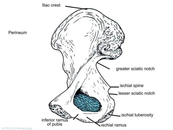
Boundaries & Division of Perineum Boundaries: • Its bony boundaries are: • Anterior:Symphysis pubis. • Posterior:Coccyx. • Lateral:Ischiopubic rami, ischialtuberosities & sacrotuberous ligaments. • Division: • By an imaginary line passing through two ischialtuberosities, it is divided into: • Urogenital triangle anteriorly. • Anal triangle posteriorly.
Urogenital Triangle Boundaries : Anteriorly : Symphysis pubis Posteriorly : Transverse line passing through the 2 ischialtuberosities. Laterally :Ischiopubic rami & ischialtuberosities. Contents : • Lower part of urethra & vagina. • External genitalia (vulva). Urethra Vagina Vulva
Urogenital Diaphragm • A triangular musculofascial diaphragm located inthe anterior part of the perineum. • Fills in the gap between the pubic arch. • Composed of: Sphincter urethrae and the deep transverse perineal musclesenclosed within the superior and inferior layers of fascia of the urogenital diaphragm • The inferior layer of the fascia is formed by theperineal membrane
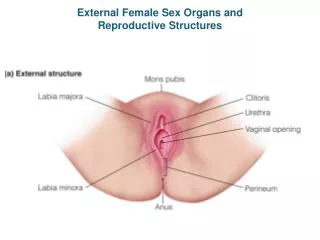
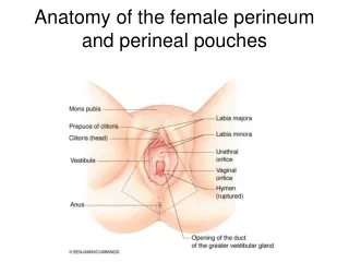
Female External Genitalia (Vulva) • Mons pubis : a collection of fat overlying the pubes. • Labia majora. • Labia minora. • Clitoris. • Vestibule of vagina: The interval between the two labia minora. • Vagina & urethra open into the vestibule through urethral orifice anteriorly and vaginal orifice posteriorly.
Fascia of Urogenital Triangle (Perineal Fascia) The perineal fascia is continuous anteriorly with the fascia of abdomen and consists of superficial and deep layers Superficial perineal fascia: consists of: Superficial fatty layer (Camper’s fascia) makes up the substance of mons pubis & labia majoraand extends into the anal region Deep membranous layer (Colle’s fascia ): Does not extend to anal region. Becomes fused with the posterior margin of the perineal membrane Deep perineal fascia invests the muscles in the superficial perineal pouch
It is the space between the deep membranous layer of superficial fascia and the perineal membrane. BOUNDARIES: Inferiorly: membranous layer of superficial fascia. Superiorly: perineal membrane. Laterally:ischiopubicrami Superficial Perineal Pouch
Contents of Superficial Perineal Pouch Bulbs of vestibule:on each side of vaginal orifice. Crura of clitoris. Superficial perineal muscles: Bulbospongiosus muscle, surrounds orifice of vagina and coversvestibular bulb. Ischiocavernosus muscle, covers crus of clitoris on each side. Superficial transverse perinealmuscles. Greater vestibular glands: on each side of vaginal orifice. Perineal branch of pudendal nerve supplying muscles & skin.
Deep Perineal Pouch It is a completely closed space deep to the perineal membrane BOUNDARIES: Inferiorly:Inferior fascia of the urogenital diaphragm (Perineal membrane) Superiorly: Superior fascia of the urogenital diaphragm Laterally: Inferior portion of obturatorinternusfacia. Coronal section of pelvis
Part of urethra Part of vagina Sphincter urethrae muscle, which is pierced by urethra & vagina. Deep transverse perineal muscles Internal pudendal vessels Dosal nerve of clitoris Contents of Deep Perineal Pouch
Vagina • The vagina is a muscular canal that leads from the uterus to the external orifice of the genital canal • It measures about 3 in. (8 cm) long. • It serves as the excretory duct for the menstrual flow & forms part of the birth canal. • The vaginal orifice in a virgin possesses a thin mucosal fold, called the hymen, which is perforated at its center. • Arteries: • Vaginal artery, a branch of the internal iliac artery • Vaginal branch of the uterine artery • Veins: drain into the internal iliac veins.
Anal Triangle Boundaries: Anteriorly: Transverse line passing through the 2 ischialtuberosities. Posteriorly : coccyx. Laterally :ischialtuberosity & sacrotuberouslig. Contents: Lower part of Anal canal Ano-coccygeal body Ischiorectal fossa on each side
Anal Canal • It is about 1.5 in. long, descending from the rectal ampullato the anus. • Relations(In female): • Anteriorly:Perineal body, urogenital diaphragm, and lower part of vagina • Posteriorly:Anococcygeal body. • Laterally:Ischiorectalfossae. • Division:Divided into: • Upper half: derived from hindgut (endoderm) • Lower half: derived from the proctodeum (ectoderm) • The two parts have different blood supply,nerve supply and lymphatic drainage.
IschiorectalFossa • A fascial lined wedge-shaped space on each side of the anal canal. Boundaries: • Base: Skin of the perineum. • Medial wall: Levatorani & anal canal. • Lateral wall: Obturatorinternus, covered with fascia. Contents: • Dense fat. • Pudendal nerve & internal pudendal vesselswithin the pudendal canal • Inferior rectal nerve & vesselscrossing the fossa to reach anal canal.
Pudendal Canal: • A fascial canal formed by obturator fascia, located on the lateral wall of the ischiorectalfossa • Contents: • Pudendal nerve. • Internal pudendal vessels.
Pudendal Nerve Block Pudendal nerve block is used in providing analgesia for the second stage of labourand to provide anesthesia of the perineum in order to create or repair an episiotomy. Can be done by transvaginallyor through perineal approach. Transvaginal method:The needle is passed through the vaginal mucous membrane toward the ischial spine. After the needle is passed through the sacrospinous ligament, the anesthetic solution is injected around the pudendal nerve Perineal method: The ischialtuberosityis palpatedsubcutaneously through the buttock. The needle is inserted on the medial side of the ischialtuberosityto a depth of about 1 in. (2.5 cm) from the free surface of the tuberosity. The anesthetic is injected around the pudendal nerve. An episiotomy is a surgically planned incision on the perineum and the posterior vaginal wall during second stage of labor to prevent perineal tear.
Thank You & Good Luck