Download
1 / 17
490 likes | 4.84k Views
Extrachromosomal Replicons. Chapter 16. 16.2 The Ends of Linear DNA Are a Problem for Replication. Special arrangements must be made to replicate the DNA strand with a 5′ end. Figure 16.1. 16.3 Terminal Proteins Enable Initiation at the Ends of Viral DNAs. A terminal protein:

E N D
Extrachromosomal Replicons Chapter 16
16.2 The Ends of Linear DNA Are a Problem for Replication • Special arrangements must be made to replicate the DNA strand with a 5′ end. Figure 16.1
16.3 Terminal Proteins Enable Initiation at the Ends of Viral DNAs • A terminal protein: • binds to the 5′ end of DNA • provides a cytidine nucleotide with a 3′–OH end that primes replication Figure 16.4
16.4 Rolling Circles Produce Multimers of a Replicon • A rolling circle generates single-stranded multimers of the original sequence. Figure 16.5
16.5 Rolling Circles Are Used to Replicate Phage Genomes • The ϕX A protein is a cis-acting relaxase. • It generates single-stranded circles from the tail produced by rolling circle replication. Figure 16.8
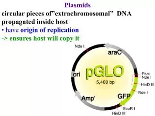
16.6 The F Plasmid Is Transferred by Conjugation between Bacteria • A free F factor is a replicon that is maintained at the level of one plasmid per bacterial chromosome. • An F factor can integrate into the bacterial chromosome • Its own replication system is suppressed. • The F factor codes for specific pili that form on the surface of the bacterium. Figure 16.9
An F-pilus enables an F-positive bacterium to: • contact an F-negative bacterium • initiate conjugation Figure 16.10
16.7 Conjugation Transfers Single-Stranded DNA • Transfer of an F factor is initiated when rolling circle replication begins at oriT. • The free 5′ end initiates transfer into the recipient bacterium. • The transferred DNA is converted into double-stranded form in the recipient bacterium. Figure 16.11
When an F factor is free, conjugation “infects” the recipient bacterium with a copy of the F factor. • When an F factor is integrated, conjugation causes transfer of the bacterial chromosome. • Transfer continues until the process is interrupted by (random) breakage of the contact between donor and recipient bacteria. Figure 16.12
16.8 The Bacterial Ti Plasmid Causes Crown Gall Disease in Plants • Infection with the bacterium A. tumefaciens can transform plant cells into tumors. • The infectious agent is a plasmid carried by the bacterium. Figure 16.13
The plasmid also carries genes for synthesizing and metabolizing opines (arginine derivatives) • They are used by the tumor cell. Figure 16.14
16.9 T-DNA Carries Genes Required for Infection • Part of the DNA of the Ti plasmid is transferred to the plant cell nucleus. Figure 16.15
The vir genes of the Ti plasmid are: • located outside the transferred region • required for the transfer process Figure 16.17
The vir genes are induced by phenolic compounds released by plants in response to wounding. Figure 16.18
The membrane protein VirA is autophosphorylated on histidine when it binds an inducer. • VirA activates VirG by transferring the phosphate group to it. • The VirA-VirG is one of several bacterial two component systems that use a phosphohistidine relay. Figure 16.19
16.10 Transfer of T-DNA Resembles Bacterial Conjugation • T-DNA is generated when a nick at the right boundary creates a primer for synthesis of a new DNA strand. • The preexisting single strand that is displaced by the new synthesis is transferred to the plant cell nucleus. • Transfer is terminated when DNA synthesis reaches a nick at the left boundary.
The T-DNA is transferred as a complex of single-stranded DNA with the VirE2 single strand-binding protein. • The single stranded T-DNA is: • converted into double stranded DNA • integrated into the plant genome • The mechanism of integration is not known. • T-DNA can be used to transfer genes into a plant nucleus.