Download
1 / 30
300 likes | 314 Views
Understand the structure and composition of bones and cartilage, their types, classifications, and key roles in the skeletal system. Explore the vital functions of the skull, spine, mandible, and other crucial skeletal components.

E N D
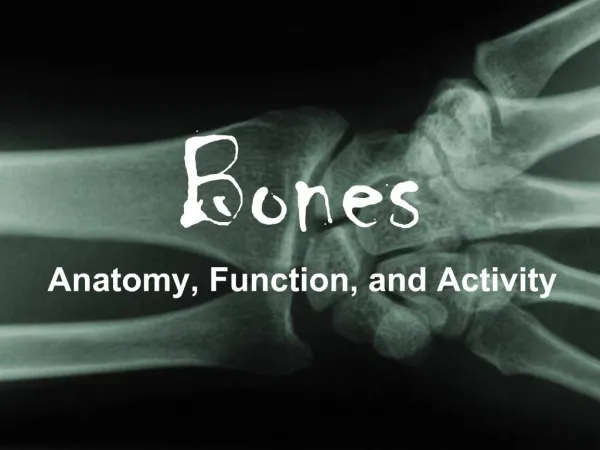
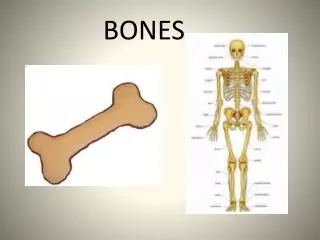
Bones Miss Ulrich
Cartilage • Connective tissue • Most of the skeleton is initially formed by fast-growing cartilage. This is replaced by bone tissue in the fetal and childhood periods • Consists primarily of water (60-80%) and is very resilient --> has the ability to spring back to original shape after being compressed
Types of Cartilage • Hyaline: most common form of cartilage • Provides support through flexibility and resilience. • Makes up articular cartilage (covers the ends of adjoining bones in movable joints • Also forms the cartilaginous attachments of the ribs to sternum. • Most of the cartilage found in the respiratory structures and the embryonic skeleton
Types of Cartilage • Elastic: similar to hyaline but more elastic • Better to tolerate repeated bending • Epiglottis is made up of elastic cartilage • Fibrocartilage: unusual tissue that resist both strong compression and strong tension (pulling) forces. • In certain ligaments (i.e., discs between vertebrae and menisci of the knee)
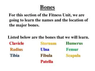
Classification of Bones Bone are classified by their shape: • Long bones: most bones in the limbs are long bones • Short bones: roughly cubed shaped. Occur in wrist and ankle • Flat bones: thin, flattened, and usually somewhat curved. Most cranial bones, ribs, sternum, scapula • Irregular bones: various shapes and do not fit into other categories . Hip bones, vertebrae.
The Skull • a bony structure • Support the structure of the face and creates a cavity for the brain • provides attachments for the head and neck muscles • At birth, the skull is large in comparison to the rest of the body, and a baby's skull is compressible. The "soft spots" in a baby's head harden and grow together until the bones meet and mesh like a jigsaw puzzle. • The largest of the six main soft spots is a diamond-shaped area near the middle of the top of the skull. This is the last area to harden and close, usually at about the age of eighteen months.
The Mandible • Largest, strongest bone of the face • Our Jaw • Serves for the reception of the lower teeth • Temporomandibular Joint (TMJ) is the joint that acts as a hinge as well as gliding mechanism
The SpineThethree types of vertebra • Cervical vertebrae contain three holes • Located in the neck • Thoracic vertebrae have a long, thin spinous process that is angled downward (it looks like a giraffe) • The lumbar vertebrae have a short spinous process that is flat and sticks straight out (it looks like a moose)
The sacrum and coccyx bones • The sacrum is a triangular shaped bone with many openings for nerves • It is located in the back of the pelvis • Between the red brackets • The coccyx bone is the tailbone that hangs down from the sacrum • The blue arrows point to the coccyx
Where are the back bones located on the body? • The cervical is in the neck • Fracture often leads to paralysis or death • The thoracic is in the upper back • Most common fracture of the spine along with lumbar • The lumbar is in the lower back • Most common fracture of the spine along with thoracic • Severe back pain, if spinal cord involved: numbness, tingling, weakness, bowel/bladder dystfunction • The sacrum is below the lumbar vertebra • Low back pain, buttock, or hip pain, pain in the groin and front portion of thigh. Weakness is lower limbs, bladder control • The coccyx hangs off the sacrum • Common from falls. More common in females due to wider pelvis cervical thoracic lumbar sacrum coccyx
The clavicle • The clavicle is the only ‘s’ shaped bone in the body…it is our collar bone • Acts as braces (hold scapulae and arms out laterally from thorax) • Commonly fractured. Usually by a fall • Signs and Symptoms: • Sharp pain with movement • Swelling • Referred pain • Possible nausea, dizziness • Treatment: rest and sling clavicle
The Scapula • The scapula is your shoulder blade • The scapula contains a large ridge on the posterior side that is referred to as the scapular spine (the red arrow points to the scapular spine) • Fracture is caused by blunt trauma • Symptoms: • Extreme pain when moving arm • Swelling around back of shoulder • Skin abrasions • Treatment: sling works for most fractures. Sometimes hospitalized
The Sternum • The sternum is the breastbone • The sternum is composed of three bones (specifically) • The manubrium is the enlarged upper section • The body is the blade-like center section • The xiphoid process is the downward projection from the body of the sternum • Fracture caused by blunt trauma • Symptoms: • Pain, tenderness, bruising, swelling, may be bent or deformed • Treatment: reducing pain and limiting movement. Fractures often come with internal injuries
The Ribs • A thin curved bone • The body has 12 pairs of ribs • All attach to the thoracic vertebrae posteriorly and run to the front of the chest • The top 7 pairs attach directly to the sternum and are called true ribs • The bottom 5 pairs attach to the sternum either indirectly or not at all and are called false ribs • Middle ribs are most commonly fractured, usually from direct blows. The 7th and 10th rib are most commonly fractured. • Symptoms: pain/ grating sound while breathing or with movement
The Humerus • The humerus is the upper arm bone • It has a large round knob at the proximal end which fits into the cavity of the scapula • This creates a “ball and socket joint” • Fracture symptoms: severe bone pain, swelling, bruising, tenderness • Treatment: hanging cast, surgery
The Ulna • The ulna is a forearm bone that has a ‘u’ shape at the proximal end • Red arrow indicates the bone • Slightly longer than the radius • Main bone forming the elbow joint with the humerus • Looks like a monkey wrench • Causes of fractures: sporting accident, repetitive stress • Symptoms: pain, swelling, arm deformity tenderness
The Radius • The radius is the forearm bone that has a ‘golf tee’ like dip in the proximal end • Rotates over the ulna when pronating • Most common fracture in the arm • Casting, surgery
Carpals, metacarpals and phalanges of the hand (general) • The carpals are the wrist bones • The metacarpals are the bones of the palm of the hand • The phalanges of the hand are the finger bones • These bones will be asked on articulated hands (assembled together) • Most injuries result from direct trauma • Treatment: casting, splints, pins, metal plates/screws carpals metacarpals phalanges
The Coxal bone • The coxal bone is the hip bone. • Three bones make up the coxal • Ischium is the rough thick section, it is what you sit on (green color) • Ilium is the large, flat upper section (cream color) • Pubis is the thin, curved anterior, inferior section of the coxal bone (orange color) • Fractures are difficult to heal. 90% are in patients 65 or over.
The Femur • The femur is the thigh bone • Largest, longest, and strongest bone of the body and highly vascular • The femur has a round knob on the proximal end on the medial side • The main shaft of the bone bows forward • Most fractures are treated surgically • Up to 40% require blood transfusion
The Patella • Knee cap (looks like a little rock) • Protects the anterior articular surface of the knee joint • Functional role: knee extension • Increases the leverage that the tendon can exert on the femur by increasing the angle at which it acts • Dislocations occur with significant regularity, particularly in young female athletes • Patella slides out of position, with intense pain and swelling • Can be tracked back into place with extension of the knee
The Tibia • The large bone of the lower leg, the proximal end is more enlarged • Weight baring • Medial malleolus is the medial projection at the distal end • Shaft fractures are most common • Cast is most common treatment, rodding may also take place
The Fibula • Smaller, thinner bone of the lower leg • The lateral malleolus is the lateral projection at the distal end of the bone • Most common fracture comes from an rolling your ankle inwards
The FootTarsals, metatarsals, and phalanges (general) • Tarsals are the ‘ankle bones’ • Metatarsals are the bones of the flat part of the food • Phalanges of the foot are the bones of the toes • Fractures in the foot are very common tarsals metatarsals phalanges
Disorders of Bones Osteoporosis • The most common type of bone disease. (1 out of 5 women over the age of 50) • Occurs when the body fails to form enough new bone, when too much old bone is reabsorbed by the body, or both. • Calcium and Phosphate are two minerals that are essential for normal bone formation
Disorders of Bones (cont’d) • As you age, calcium and phosphate may be reabsorbed back into the body from the bones, which makes tissue weaker. • Results in: brittle, fragile bones that are prone to fractures. Even without injury • Loss usually occurs gradually over the years. • Leading cause are a drop in estrogen in women at the time of menopause and a drop in testosterone in men • Higher risk in women over the age of 50 and men over the age of 70 Testing: bone mineral density test, spine CT, spine or hip x-ray
Disorders of Bones (cont’d) Osteosarcoma • Cancerous bone tumor • Develops during the period of rapid growth that occurs in adolescence. • Average age at diagnosis is 15 (between the age of 10-25) • Males and females have similar incidence until late adolescence, when males are more commonly affected.
Disorders of Bones (cont’d) • Cause is unknown. • Commonly occurs in the bones of the: • Shin (near the knee) • Thigh (near the near) • Upper arm (near the shoulder) • In large bones and in the area of bone with fastest growth rate • Complications: • Limb removal • Spread of cancer to the lungs • Side effects of chemotherapy Testing: biopsy, blood tests, bone scan, CT scan, x-ray)
Disorders of Bones (cont’d) Scoliosis • The “twisted disease” • 3 general causes: • Congenital (present at birth)- due to a problem with the formation of the vertebrae or fused ribs during development in the womb or in early life • Neuromuscular: poor muscle control or muscle weakness, or paralysis due to diseases such as cerebral palsy, muscular dystrophy, etc. • Idiopathic scoliosis- unknown cause. Most common in adolescents • Most cases occur is girls
Disorders of Bones (cont’d) Scoliosis • Symptoms: • One shoulder appears to be higher than the other • Pelvis appears to be tilted • Backache or low-back pain • Fatigue • Shoulders or hips appear uneven • Spine curves abnormally to the side (laterally) Testing: Scoliometer screening (measure curvature of the spine), spine X-rays, MRI)