Download
1 / 22
230 likes | 417 Views
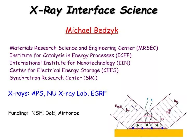
X -Ray Interface Science. Michael Bedzyk Materials Research Science and Engineering Center ( MRSEC ) Institute for Catalysis in Energy Processes (ICEP ) International Institute for Nanotechnology (IIN) Center for Electrical Energy Storage (CEES) Synchrotron Research Center (SRC).

E N D
X-Ray Interface Science Michael Bedzyk Materials Research Science and Engineering Center (MRSEC) Institute for Catalysis in Energy Processes (ICEP) International Institute for Nanotechnology (IIN) Center for Electrical Energy Storage (CEES) Synchrotron Research Center (SRC) X-rays: APS, NU X-ray Lab, ESRF Funding: NSF, DoE, Airforce
Group Party June 2013 Group breakdown: 2 postdocs, 7 graduate students
Bedzyk Group Overview: Atomic Scale View of Interfacial and Nanoscale Processes with X-Rays Nanoscale Electrodes for Li-Ion Batteries X-ray Standing Wave studies of graphene A10 18bp duplex Au X-ray Scattering and Absorption Studies of Au Nanostructures for DNA Functionalization and Assembly C3-SH Ion distribution around DNA-NPs DNA-NP Schematic Nanorod growth and functionalization
Some X-ray Basics:Wave Property Structural Infoλ= 0.1 to 10 Å wavelength E-M radiation X-rays scatter coherently from electronsParticle Property Compositional InfoEϒ = 1 to 100 keV energy Photo effect: Inner shell (K, L) ionization XRF : Decay of excited ion to ground state by characteristic XRF emission
X-ray Vision Advantage: Weak interaction with matter High penetrating power In situ analysis Buried structures Atomic-scale resolution Problem: Weak interaction with matter weak signal Need very intense X-ray source
Brightest X-ray Source in Western Hemisphere = Advanced Photon Source Undulator Device relativistic electrons pass thru periodic magnetic array
Argonne National Laboratory Funded by US Dept. of Energy Lab NU NU-ANL Carpool ORD ANL
Simultaneous SAXS-MAXS-WAXS at DND-CAT/APS 3 CCD Areal Detectors MAXS Capillary Tube with flowing Sample Solution SAXS WAXS Incident X-ray Beam $1.2 M, Just completed Upgrade
Self-assembled systems of amphiphiles A Critical packing parameter = V/AL Spherical micelle V L Fiber hydrophilic Curved membrane hydrophobic Planar membrane
Applications Drug delivery Cell model Photovoltaic cells Gene therapy Template for synthesis, tissue regeneration…..
Shells of different shapes HIV virus (~150 nm across) Mimvirus (~200 nm across) Mouse Polyoma Virus (~50 nm) icosahedral spherical (Dubois, et al., Nature 2001) Crystalline lipid vesicle (~1 mm across)
100 nm -size and shape variability of cellular carboxysomes (Iancu, et al., J. Mol. Biol. (2010) 396, 105–117) -Walby’s archaea organism -hexagonal lattice (W. Stoeckenius J. BACTERIOLOGY, (1981)) - Mixed component system
- Fluid Membranes (no internal order): Young’s modulus (Y) = 0 Bending rigidity (κ) Catanionic self-assembled membranes anion cation - + + cones cylinders - Crystalline membranes (with internal order): Young’s modulus > 0
Quick-freeze deep-etch TEM microscopy images Cation alone Cation + anion mixture 100nm 500 nm 500 nm
SAXS - 1-100 nm scale features - size and shape I WAXS - molecular packing - crystal structure q (nm-1) Small/ Wide Angle X-ray Scattering (SAXS/ WAXS) X-ray 2q Fourier Transform
SAXS/WAXS Data Processing X-Ray Vesicles or membranes flowing freely in solution 1D graph of intensity vsq Do an angle averaged integration q (Å-1) 2D images from SAXS
Model fit of bilayer structure Porod Power Law +3 Cation and -1 anion mixture vesicles α = 2 2D platelet cation Cationonly 3.8 nm 2.1 nm 5.3 nm Fit the data with a bilayer model to obtain thickness
Molecular packing within membrane +3 Cation and -1 anion mixture vesicles Cation alone α = 2 WAXS d = 2π/q = λ/2sinθ = 0.413 nm Hexagonal lattice Electrostatic attraction induces crystallization of tails Area/ molecule = 0.197 nm2 Packing of tails 0.477 nm
Why do we want to control membrane crystal structures? - Crystal structure can change morphology - Molecule flow rate across membrane can be controlled by packing density and membrane thickness - Hydrophobic drugs encapsulated inside membrane
Questions • Can we control the crystal structure? • Can we control the shape of the vesicles or membrane morphology? • Play with electrostatics! • Change pH to change effective charge of head groups. • Change tail length to change dipolar van der Waals attraction
What a new student in the Bedzyk group might expect to be involved with while pursuing their PhD • Gain an expertise with general x-ray techniques and experimental design • Learn fundamental materials science/ chemistry/ physics/ biology relevant to the systems they are studying (interdisciplinary research) • Take measurements at the Advanced Photon Source and help develop the Dupont-Northwestern-Dow beamline (sector 5) • Understand atomic-scale structure and how it applies to desirable materials properties