Download

1 / 29
550 likes | 4.69k Views
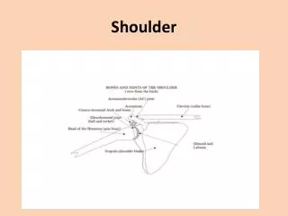
SHOULDER PRESENTATION. Dr. S.K.S. Shoulder presentation :- when the long axis of fetus lies transversely with long axis of maternal spine, as a result shoulder of fetus occupies the birth canal. Incidence: 0.3%. Position of fetus. Dorso-anterior- more common. Dorso-posterior.

E N D
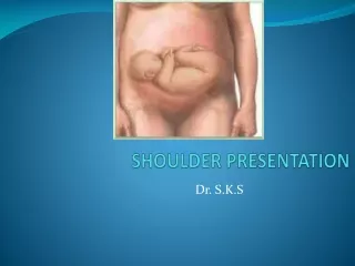
SHOULDER PRESENTATION Dr. S.K.S
Shoulder presentation :- when the long axis of fetus lies transversely with long axis of maternal spine, as a result shoulder of fetus occupies the birth canal. • Incidence: 0.3%
Position of fetus • Dorso-anterior- more common. • Dorso-posterior.
Mechanics of presentation: • long axis of the fetus is perpendicular to long axis of mother (ie occurs in transverse lie) • mostly the shoulder presents in a transverse lie, but alternative presentations are • hand and arm (may be prolapsed into the vagina) • cord • nil (fetal back is down, and above the level of the inlet)
AETIOLOGY • Fetal • prematurity, multiple • Liquor • polyhydramnios • Uterine • Anomaly esp. subseptate uterus • Placenta • praevia • Pelvis • contraction, tumour • Parity • high maternal parity (80% of cases occur in women who are para3 or more)
DIAGNOSIS • Inspection:- Asymmetrical enlargement of uterus. Abdomen is transversely broad. • Palpation Fundal ht.:- smaller than gestational age. Fundal grip:- absent. Lateral grip:- head of fetus in one side and breech on other side. Pelvic grip:- empty.
Auscultation :- FHS is heard at higher level and more distinct in dorso-anterior position • P/V examination:- During pregnancy:- high presenting part. During labour:- shoulder is identified by palpating the following parts:- acromian process, scapula, clavicle and axilla. After rupture of membrane- hand may be prolapsed.
MANAGEMENT • General management:- I/V fluid Blood grouping, Rh typing & cross match at least 1 pint blood. Parenteral antibiotics – ampicillin 1 gm, metronidazole 500mg.
Obstetric management :- • Antenatal management • Labour management.
A. Antenatal management External cephalic version if not contraindicated at 32-34 wks. If fail repeat after 1 week Vertex presentation If revert back to transverse lie NVD El. C/S.
B. Labour management Labour Dead baby Alive baby Internal podalic version with breech extraction C/S Destructive operation C/S
Immediate C/S must be perform if:- • Cord prolapse • Early rupture of membrane • ECV failed. • Any delay in the progress of labour.
Danger of transverse lie • Maternal • Prolong labour • Obstructed labour • Rupture of uterus • Haemorrhage & shock • Maternal death • Fetal • Cord prolapse • Hand prolapse • IUD • Foetal distress • Still birth
COMPOUND PRESENTATION When more than 1 presenting part enters birth canal at a time or, When a fetal extremity prolapses alongside the presenting part, and both enter the maternal pelvis at the same time • vertex-hand or cord • breech-hand or cord • vertex-arm-foot • Incidence: 0.1% • Aetiology • Fetal • multiple • premature • Maternal • Multiparity
MANGEMENT • Exclude cord prolapse • occurs in up to 20% of cases • Otherwise expectant • vertex-foot: try to gently reposition the lower extremity • if arm prolapses in vertex-hand - deliver by CS
CORD PROLAPSE/PRESENTATION Def – when the umbilical cord descends along with the presenting part, it is called cord prolapse/presentation. Clinically, it can be divided as – Occult prolapse – cord remains by the side of the presenting part & is not felt . Cord presentation – cord is slipped down below the presenting part. Cord prolapse – cord is lying inside the vagina or outside the vulva following rupture of membranes
Incidence – 1 in 300 deliveries. Mostly found in parous women. Etiology – following factors play a great role. • Malpresentation. • Contracted pelvis • Pre maturity. • Twins. • Hydramnios • Long cord • Iatrogenic – low rupture of membrane, rotation / version.
Diagnosis – Occult prolapse – difficult to diagnose. Cord presentation – by feeling the pulsation of cord. Cord prolapse – cord can be felt pulsating if the fetus is alive. Cord pulsation may cease during uterine contraction but returns soon after contraction passes off. Fetus may be alive even in the absence of cord pulsation, hence USG helps determine cardiac movt.
Management – • Once the diagnosis is made, try to preserve the membranes & to expedite the delivery. • If immediate vaginal delivery is not possible or contraindicated, caesarean section is the best choice. • Management Aim is guided by – a. baby living or dead. b. maturity of the baby. c. dilatation of the cervix.
Baby living – -i.v. fluids & oxygen by mask. -Bladder filling to be done to raise the presenting part, 400-750 ml of NS is used with a Foleys catheter, the balloon is inflated & catheter is clamped. Empty the bladder before CS. - lift the presenting part off the cord. - keep the pt. in sims position. - to replace the cord inside the vagina (to minimize vasospasm due to irritation). - caesarean section is the best treatment when the baby is viable.
Immediate safe vaginal delivery is possible if the head is engaged. Immediate delivery to be completed by forceps. If breech – by breech extraction. Baby dead – labour should be allowed to proceed. No need ofCS.
Prognosis – Fetal – fetus is at greater risk of anoxia. The hazards to the fetus is more in vertex presentation. The perinatal mortality is about 50%. Maternal – operative delivery risks of anesthesia, blood loss & infection.