Download
1 / 11
110 likes | 332 Views
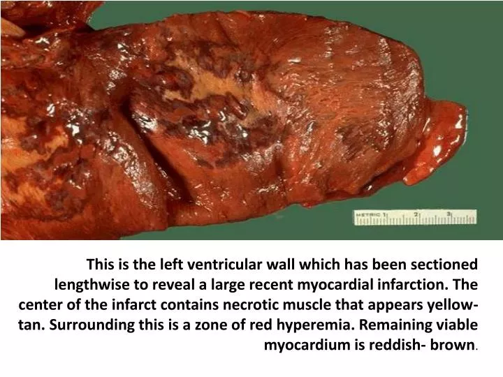
This is the left ventricular wall which has been sectioned lengthwise to reveal a large recent myocardial infarction. The center of the infarct contains necrotic muscle that appears yellow-tan. Surrounding this is a zone of red hyperemia. Remaining viable myocardium is reddish- brown .

E N D
This is the left ventricular wall which has been sectioned lengthwise to reveal a large recent myocardial infarction. The center of the infarct contains necrotic muscle that appears yellow-tan. Surrounding this is a zone of red hyperemia. Remaining viable myocardium is reddish- brown.
The heart is opened to reveal the left ventricular free wall on the right and the septum in the center. There has been a remote myocardial infarction that extensively involved the anterior left ventricular free wall and septum.
There is a tear (arrow) located 7 cm above the aortic valve and proximal to the great vessels in this aorta with marked atherosclerosis. This is an aortic dissection.
In the region of the foramen ovale on the interatrial septum is a small atrialseptal defect, as seen in this heart opened on the right side. Here the defect is not closed by the septum secundum, so a shunt exists across from left to right.
This left ventricle is very thickened (slightly over 2 cm in thickness), but the rest of the heart is not greatly enlarged. This is typical for hypertensive heart disease. The hypertension creates a greater pressure load on the heart to induce the hypertrophy.
The dark red infarcted small intestine contrasts with the light pink viable bowel. The forceps extend through an internal hernia in which a loop of bowel and mesentery has been caught. This is one complication of adhesions from previous surgery. The trapped bowel has lost its blood supply.
The white arrow denotes the most prominent fatty streak in the photo, but there are other fatty streaks scattered over the aortic surface. Fatty streaks are the earliest lesions seen with atherosclerosis in arteries.
These three aortas demonstrate mild, moderate, and severe atherosclerosis from bottom to top. At the bottom, the mild atherosclerosis shows only scattered lipid plaques. The aorta in the middle shows many more larger plaques. The severe atherosclerosis in the aorta at the top shows extensive ulceration in the plaques.
The pericarditis here not only has fibrin, but also hemorrhage. Thus, this is called a "hemorrhagic pericarditis". It is really just fibrinouspericarditis with hemorrhage. Without inflammation, blood in the pericardial sac would be called "hemopericardium".
Jars 32/14---------------------------------------------BOWEL INFARCT 21/17----------------------------------EARLY ATHEROSCLEROSIS 27/17---------------------------ADVANCED ATHEROSCLEROSIS 37/17-------------------------- ADVANCED ATHEROSCLEROSIS 46/17---------------------------------------AORTIC DISSECTION 25/17---------------------LEFT VENTRICULAR HYPERTROPHY 43/17--------------------------------ARTIFICIAL MITRAL VALVE 22/18 ----- ------------------------------------ LUNG INFARCT 4/17------------------------OLD MYOCARDIAL INFARCTION 45/17-------------------------------ARTERIAL SEPTAL DEFECT 6/17----------------------------ACUTE PERICARDITIS(CHILD) 47/17-------------------RECENT MYOCARDIAL INFARCTION