Download
1 / 50
540 likes | 912 Views
Chromosomes & Cell Cycle SDK October 1, 2013. Road Map. Chromosomes Chromosome Anatomy Defining Chromosomal Location Cell Cycle Cell Division Mitosis Phases of Mitosis Cell Cycle Checkpoints Functions of Cell Division Uncontrolled Cell division(Cancer) Sexual Reproduction

E N D
Road Map • Chromosomes • Chromosome Anatomy • Defining Chromosomal Location • Cell Cycle • Cell Division • Mitosis • Phases of Mitosis • Cell Cycle Checkpoints • Functions of Cell Division • Uncontrolled Cell division(Cancer) • Sexual Reproduction • Sex Cells – Gametes • Meosis • Significance of Meiosis • Mitosis vs Meiosis • KARYOTYPE • Making a Karyotype • Individual chromosomal importance • Books
Chromosomes • Linear units of DNA • Thread-like structures located inside the nucleus, • Each chromosome is made of protein and a molecule of deoxyribonucleic acid (DNA). • Come in pairs (homologous pairs) • Each species has characteristic number • Human has 23 pairs of the chromosome • 22 are the autosomes • One is Sex Chromosome
It is a combination of two words, i.e., “Chroma”-means ‘colour’ and “Somes”-means ‘body’. So the coloured thread like bodies present in the nucleus of the living cells, which helps in the inheritance (transmission) of characters in form of Genes from generation to generation are known as CHROMOSOMES. CHROMOSOME
Chromosome Anatomy • Chromosomes come in two forms: • Unduplicated(Monad state) • Duplicated(Diads state) • Duplicated chromosomes are made up of two chromatids • Referred to as sister chromatids • Chromatids are joined at a region called the Centromere • Chromatids are identical • Each chromatid is a double helix of DNA • The diad state is what is typically seen in pictures of chromosomes, but it only occurs briefly in the life of the cell. During most of interphase
Chromosome Anatomy • Each chromosome has two components • Telomere (Upper & Lower end of chromosomes) • Constriction Point/Centromere (Position is variable) • The constriction point called the centromere, which divides the chromosome into two sections, or “arms” A short arm and A long arm. • The short arm of the chromosome is labeled the “p arm” The long arm of the chromosome is labeled the “q arm” .
Centromere • Sometimes called the “primary constriction” on a chromosome • This is the attachment point for the spindle • This is the many repeats of about 170 bp element. • The location of the centromere on each chromosome gives the chromosome its characteristic shape, and can be used to help describe the location of specific genes and further classify the chromosomes.
Types of Chromosomes There are four types of chromosomes based upon the position of the centromere. 1) Metacentric : Centromere occurs in the centre and all the four chromatids are of equal length. 2) Submetacentric : Centromere is a little away from the centre and therefore chromatids of one side are slightly longer than the other side. 3) Acrocentric : Centromere is located closer to one end of chromatid therefore the chromatids on opposite side are very long. A small round structure, attached by a very thin thread is observed on the side of shorter chromatid. The small round structure that is a part of the chromatid is termed as satellite. 4) Telocentric : Centromere is placed at one end of the chromatid and hence only one arm. Such telocentric chromosomes are not seen in human cells.
Defining Chromosomal Location • Each chromosome is further divided into regions, labeled p1,p2,p3…. and q1,q2,q3…. counting outwards from the centromere. • Regions are divided into bands and sub-bands labeled p11.1, 012.3, p13.5 ….)
What are telomeres? Centromeres/Telomeres What are entromeres? The constricted region of linear chromosomes is known as the centromere. Although this constriction is called the centromere, it usually is not located exactly in the center of the chromosome and, in some cases, is located almost at the chromosome's end. Telomeres are repetitive stretches of DNA located at the ends of linear chromosomes. Teleomersconsist of as many as 2000 repeats of the sequence: 5' TTAGGG 3'. They protect the ends of chromosomes in a manner similar to the way the tips of shoelaces keep them from unraveling.
The normal human chromosome diploid number is: • 23 • 24 • 46 • 48 46
The chromosome which picture is shown below is 1. Metacentric 2) Submetacentric 3) Acrocentric 4) Telocentric Submetacentric
The mark point on the Chromosome is • P arm • q arm • Centromere • Sub band Centromere
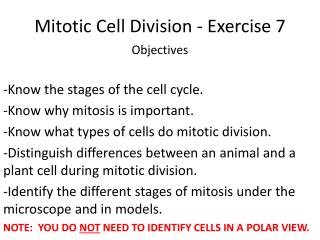
Cell Cycle • The continuity of life is based upon the reproduction of cells, or cell division • A typical human cell undergoes a division about every 24 hours (there are many exceptions!) • The cell cycle is basically an alternation of 2 major phases – Mitosis and Interphase • Interphase is the phase in which the cell spends 23 of the 24 hours • The cell grows and preform its normal cell activity • Mitosis takes about 1 hour & cell division phase
The Cell Cycle • Interphase can be broken down into 3 distinct sub-phases: • G1 (Known as gap 1) • S (for synthesis of DNA) • G2 (gap 2) • Cells that do not divide are considered to be in a G0 phase – where they carry on normal housekeeping and do not prepare to divide. • Mitosis. Is comprised of Prophase, Anaphase, Metaphase and Telophase.
Cell Division • Cell division is of two types • Mitosis • Meosis • Mitosis is a process of cell duplication, or reproduction, during which one cell gives rise to two genetically identical daughter cells. Take place in all somatic cells • Meiosis, is a division of a germ cell( sperm/ova) involving two fissions of the nucleus and giving rise to four gametes, or sex cells, each possessing half the number of chromosomes of the original cell. Take place only in germ cells.
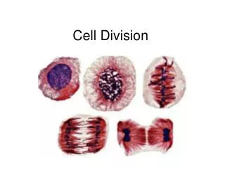
Mitosis Mitosis is ordinary cell division among the cells of the body. During mitosis the chromosomes are divided evenly, so that each of the two daughter cells ends up with 1 copy of each chromosome. • In mitosis the homologs chromosomedo not pair up. • Rather they behave independently. • Each resultant cell receives one copy of each homolog.
Phases of Mitosis • Prophase: • Chromosomes condense • Nuclear envelope disappears • Chromosome is visible • Centrioles move to opposite ends of the cell • Spindle forms • Metaphase: Chromosomes are lined up on cell equator, attached to the spindle at the centromeres
Mitosis 3. Anaphase: • Centromeres divide. Now chromosomes are monads • The monad chromosomes are pulled to opposite poles by the spindle. 4. Telophase: • Cytokinesis: cytoplasm divided into 2 separate cells • Chromosomes de-condense • Nuclear envelope re-forms • Spindle vanishes
Phases of Mitosis G2 of Interphase Metaphase Prometaphase Prophase Anaphase Telophase & beginning of cytokinesis Completion of cytokinesis
DNA replication takes place during • S - phase • G2 - phase • G1 - phase • Prophase • S - phase
The stage in which daughter chromosomes move toward the poles of the spindle is • Anaphase • Metaphase • Prophase • Telophase Anaphase
During which stage the chromosomes first become visible • Anaphase • Metaphase • Prophase • Telophase C. Prophase
Cell Cycle Checkpoints • If cell size inadequate G1 or G2 arrest • If nutrient supply inadequate G1 arrest • If an essential external stimulus is lacking G1 arrest (at R) • If the DNA is not replicated S arrest • If DNA damage is detected G1 or G2 arrest • If the spindle formation is improper, chromosome misalignment • M-phase arrest
(c) Tissue renewal. These dividing bone marrow cells (arrow) will give rise to new blood cells (LM). (b) Growth and development. This micrograph shows a sand dollar embryo shortly after the fertilized egg divided, forming two cells (LM). (a) Reproduction. An amoeba, a single-celled eukaryote, is dividing into two cells. Each new cell will be an individual organism (LM). Functions of Cell Division
Uncontrolled Cell divisionleads to Cancer? Uncontrolled Cell division Loss of cell cycle control and checkpoints • What does a cancerous cell look like? • Large or multiple nuclei • Irregular shape • Cells overlapping neighboring cells – loss of density-dependent or contact inhibition • Loss of anchorage dependence
Key Haploid Diploid n n Gametes n MEIOSIS FERTILIZATION Zygote 2n 2n Diploid multicellular organism Mitosis (a) Animals Sexual Reproduction • Fertilization and meiosis alternate in sexual life cycles • A life cycle is the generation-to-generation sequence of stages in the reproductive history of an organism
Sex Cells - Gametes • Unlike somatic cells, sperm and egg cells are haploid cells, containing only one set of chromosomes • At sexual maturity the ovaries and testes produce haploid gametes by meiosis • During fertilization, sperm and ovum fuse forming a diploid zygote • The zygote develops into an adult organism
Meiosis 5. Prophase of M1 is very long, with a number of sub- stages. 6.Main event in prophase of M1 is “crossing over”, also called “recombination”. 7. In crossing over, homologous chromosomes pair up, and exchange segments by breaking and rejoining at identical locations. 8. Several crossovers per chromosome, with random positions.This is the basis for linkage mapping. 9. Chromosomes that don’t recombine seem to have a high rate of non-disjunction (chromosome goes to the wrong pole). • Meiosis is the special cell division that converts diploid body cells into the haploid gametes. Only occurs in specialized cells. • Takes 2 cell divisions, M1 and M2, with no DNA synthesis between. • In humans, start with 46 chromosomes (23 pairs) in diad state. After M1, there are 2 cells with 23 diad chromosomes each. • After M2 there are 4 cells with 23 monad chromosomes each.
In meiosis the products are haploid gametes so two divisions are necessary. • Prior to the first division, the homologs pair up (synapse) and segregate from each other. In the second meiotic division sister chromatids segregate. • Each cell receives a single chromatid from only one of the two homologs.
Meiosis • Only diploid cells can divide by meiosis. • Prior to meiosis I, DNA replication occurs. • During meiosis, there will be two nuclear divisions, and the result will be four haploid nuclei. • No replication of DNA occurs between meiosis I and meiosis II.
Significance of Meiosis • Sexual Reproduction • If meiosis did not occur fusion of gametes would result in doubling of chromosomes for each successively reproduced generation • Genetic Variation • Meiosis provides opportunities for new combinations of genes to occurs in gametes via independent assortment of chromosomes (metaphase) and crossing over (prophase I). • Pairing up of homologous chromosomes- Synapsis (each pair is a bivalent, consist of 4 chromatids)
Mitosis vs Meiosis • One Division • Homologues do not pair • Centromeres divide • Each cell inherits both homologues • Mitosis is conservative producing daughter cells that are like parental cell. • Two Divisions • Homologues Pair up • In meiosis I, centromeres do not divide • Homologues segregate from each other. • Meiosis is not conservative, rather it promotes variation through segregation of chromosomes and recombination
Meiosis occurs for the human female in • Pancreas • Liver • Ovary • Kidney Ovary
In humans, at the end of the first meiotic division, the male germ cells differentiate into the • Spermatogonia • Primary spermatocytes • Secondary spermatocytes • Spermatids C. Secondary spermatocytes
Given below is a schematic break-up of the phases/stages of cell cycle Which one of the following is the correct indication of the stage/phase in the cell cycle? • Cytokinesis • Metaphase • Karyokinesis • Synthetic Phase D. Synthetic Phase
What is a Karyotype? • A picture (actual photograph) , or chromosome map, of all 46 chromosomes is called a Karyotype. • The karyotype can help identify chromosome abnormalities that are evident in either the structure or the number of chromosomes.
Homologous Pairs • Every individual has two copies of every chromosome • One from your mother • One from your father • Homologous pairs have the same genes but not always the same form of the gene • 23 pairs 23 x 2 = 46
Making a Karyotype • First the chromosomes are stained • Once stained, the chromosomes look like strings with light and dark "bands"
Making a Karyotype • Then they are organized by height and centromere location
Autosomes vs sex chromosomes • Chromosome pairs # 1- # 22 are called Autosomes • Chromosome pairs # 23 are called the X and Y chromosomes • Referred to as the Sex Chromosomes because they contain genes which determine gender • Two X’s means you are genetically female • One X and a Y means you are genetically male Presence of a Y chromosome means the person is male
Chromosome 1 Contains over 3000 genes Contains over 240 million base pairs, of which ~90% have been determined
Chromosome 2 • Contains over 2500 genes • Contains over 240 million base pairs, of which ~95% have been determined
Chromosome 4 • Contains approximately 1600 genes • Contains approximately 190 million base pairs, of which ~95% have been determined
Chromosome 7 • Contains approximately 1800 genes • Contains over 150 million base pairs, of which over 95% have been determined
Chromosome 11 • Contains approximately 2000 genes • Contains over 130 million base pairs, of which over 95% have been determined
Chromosome 13 • Contains approximately 800 genes • Contains over 110 million base pairs, of which over 80% have been determined
Chromosome 17 • Contains over 1600 genes • Contains approximately 80 million base pairs, of which over 95% have been