Download

1 / 55
560 likes | 695 Views
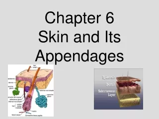
Skin and Its Appendages. Chapter 6. The Skin . The Skins Surface is as large as the body Average size Adult 17 to 20 Sq Feet Thin and relatively flat organ classified as a cutaneous membrane 2 layers that compose it Epidermis Dermis

E N D
Skin and Its Appendages Chapter 6
The Skin • The Skins Surface is as large as the body • Average size Adult 17 to 20 Sq Feet • Thin and relatively flat organ classified as a cutaneous membrane • 2 layers that compose it • Epidermis • Dermis • Forms a self-repairing and protective boundary between the internal environment of the body and the external world
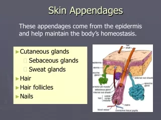
Skin • “Integument” is another name for the skin. Integumentary system is a term used to denote the skin and is appendages ( nails, hair, etc.) • 2 main layers 1) epidermis (outer and thin and avascular), 2) dermis (deeper and thick and vascular) • Beneath the dermis lies a loose subcutaneous layer rich in fat and is called the “hypodermis”. • In thick skin, the underlying dermal layer has raised papillae ridges which form finger and foot prints
Functions of Skin • Protection • Sensation – pressure, touch, temperature, pain, vibration • Excretion – regulating the volume and chemical content of sweat. • Vitamin D production – occurs when skin is exposed to ultraviolet light. • Homeostasis of body temperature
Structure of Skin • Epidermis- is the Epithelial layer that comes from the Ectodermal Layer in embryos • keratinocytes are the most important cells in the epidermis and make up 90% of the total epidermal cells • Dermis- deep layer that is relatively dense and vascular. It is connective tissue • Subcutaneous layer- rich in fat and areolar tissue ie. Hypodermis can exceed 10cm in thickness
Structure of Skin • Most of the body surface is covered by what is known as “Thin Skin” • Palms of hands, finger tips, soles of feet and other areas of friction are covered by what is known as “thick” skin • Thick Skin features • No Hair • Under lying dermal layer raised in curved ridges to form finger prints
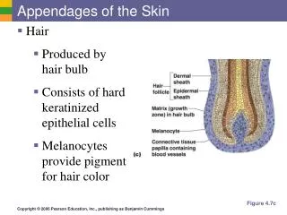
Epidermis Cell Types • Keratinocytes- become filled with tough and fibrous proteins called Keratin • Melanocytes- contribute to the color of our skin • Function is to decrease the amount of UV to the deeper layers of our skin
Cell Layers • There are typically 5 layers to the Epidermis • 1) Stratum Corneum • 2) Stratum Lucidum • 3) Stratum Granulosm • 4) Stratum Spinosum • 5) Stratum Basale
Stratum Corneum • Corneum (horny layer)-Outer most layer. A squamous layer of mostly dead cells. Tough and water repellant. Withstands wear and tear and functions as a barrier to water loss. Protects body from environmental threats, microorganisms and harmful chemicals. • Composed of dead cells where the cytoplasm has been replaced by a water repellant protein called Keratin. Continually being shed. • Hyperkeratosis- Corneum thickens far beyond normal limits= thick dry scaly skin subject to painful fissures.
Stratum Lucidum • Lucidum (clear layer) - clear layer of keratinocytes that are thickly packed together. Main function is to protect against water loss.
Strantum Granulosum/Spinosum • Granulosum (granular layer)-Cells begin to degenerate and theprocess of keratinization begins in this layer. • Spinosum (spiny layer) • 8 to 10 layers of tightly packed irregularly shaped cells • Rich in RNA to initiate protein synthesis required to make keratin
Strantum Basale • Basale (base layer) - single layer of columnar cells. They under go mitosis then migrate up through the other layers to the surface to be shed.
Epidermis Repair & Growth • The most important aspect of Integumentary system is protection. This largely depends on the ability of the epidermis to create and repair itself following injury or disease. • New cells must be formed at the same rate that old ones flake off. • Cells push up from the stratum basale through each layer to the surface. (roughly 35 days)
Epidermis Repair & Growth • The falling away of dead cells is called “desquamate” or “desquamation” • If abrasion continues over a period of time an abnormally thick stratum Corneum will develop also known as a callus. • Blisters result from injury to the cells of the epidermis or from separation of the dermal-epidermal junction. This can be any irritant that damages the physical or chemical bonds that hold the cells together ie poison ivy.
Dermal-Epidermal Junction • A specialized area between the epidermis and the dermis • Composed of a basement membrane and a polysaccharide gel that “cements” or “glues” the epidermis to the dermis.
Blisters • Result from injury to cells in the epidermis or from separation of the dermal-epidermal junction • A basic reaction of the skin to injury • Can be caused by excess friction or irritation that damages the physical or chemical bonds that hold adjacent skin cells or layers together. • Chemical agents that break disulfide linkages or hydrogen bonds cause blisters. • Desmosomes (intercellular bridges) are weakened or destroyed.
Dermis • Composed of two layers 1) papillary, 2) reticular • Much thicker than the epidermis. • This is where the mechanical strength of the skin resides. • Also provides a reservoir storage area for water and electrolytes.
Dermis • All sensory receptors for pain, touch, temperature, vibration, etc. are found in this layer. • Composed mainly of collagen and elastic fibers • Numerous muscle fibers are found here. Voluntary muscles include those responsible for facial expression. Involuntary muscles include the “arrector pili” muscles which are responsible for hair “standing on end” and for “goose pimples”
Dermal Growth & Repair • Does not continually shed • Only time of rapid regeneration is during the healing of wounds in which fibroblasts quickly reproduce forming a dense mass of new connective tissue known as a scar.
Homeostasis – Heat Regulation • 80% or more of heat transfer occurs through the skin • Heat loss can be accomplished by regulating the amount of blood flow to the skin. • If heat needs to be conserved, dermal blood vessels constrict (vasoconstriction) keeping the warmer blood circulating deeper in the body.
Homeostasis – Heat Loss • If heat loss must be increased, dermal blood vessels widen (vasodilation) increasing the skin’s supply of blood from deeper tissues. • The epidermis has 4 ways to eliminate heat • 1) Evaporation – The movement of molecules from a liquid to a gaseous state requires energy. The energy is derived from heat and causes a cooling effect. Humidity retards evaporation and therefore lessens the cooling effect and explains why the same degree of temperature seems hotter in humid climates than in dry ones.
Homeostasis – Heat Loss • 2) Radiation – the transfer of heat from the surface of one object to another without actual contact between the two. Heat radiated from the skin to cooler nearby objects. In cool environmental temperatures, radiation accounts for a greater percentage of heat loss from the skin than conduction and evaporation combined. The opposite is true in hot environments.
Homeostasis – Heat Loss • 3) Conduction – the transfer of heat to any substance actually in contact with the body. This accounts for a relatively small amount of heat loss. • 4) Convection – the transfer of heat away from a surface by movement of heated air or fluid particles. Overall, this usually accounts for very little heat loss from the body surface.
Homeostasis – Heat Production • Heat is produced by one means – metabolism of foods • Active tissues such as muscles and gland (esp. liver) produce the most heat compared to other tissues. • Chief determinant of how much heat the body produces is the amount of muscular work it does. Eg. Exercise or shivering.
Abnormal Body Temperature • Fever – known as a “febrile state”. It is associated with a systemic inflammatory response. In infections, chemical agents known as ‘pyrogens’ cause the thermostatic control centers of the hypothalamus to produce a fever. The bodies thermostat is set at a higher level causing the person to want to ‘warm up’ to the new level and produces ‘the chills’. Ultimately, the higher temperature is thought to enhance the bodies immune response. Under normal conditions, it is best to let the fever ‘break’ on its own.
Abnormal Body Temperature • Malignant Hyperthermia – an inherited condition. An abnormally increased body temperature and muscle rigidity when exposed to certain anesthetics or muscle relaxants. • Heat Exhaustion – occurs when the body loses a large amount of fluid resulting from the heat-loss mechanisms. Loss of water and electrolytes causes weakness, muscle cramps, vertigo, nausea, and possibly consciousness. Treated with rest and fluid replacement.
Abnormal Body Temperature • Heat Stroke – A.K.A. Sunstroke – severe and sometimes fatal. The inability of the body to maintain a normal temperature in an extremely warm environment. Due to old age, disease, drugs. Body temp. greater than 105 degrees. Causes rapid heart rate (tachycardia) headache, hot and dry skin, confusion, convulsions, loss of consciousness. Body must be cooled and fluids replaced immediately. A medical emergency.
Abnormal Body Temperature • Hypothermia – the inability to maintain a normal body temperature in extremely cold environment. Body temps. Less than 95 degrees. Causes shallow and slow respirations and a faint and slow pulse. Treated by slow warming of the body. • Frostbite – Damage to tissues due to extremely cold temperatures. Damage is caused by formation of ice crystals. Tissue death (necrosis) and gangrene (tissue decay) can result.
Skin Color • Dependant on the amount of melanin deposited in the cells of the epidermis. • Melanin is a dark brown pigment produced by melanocytes. It is converted from the amino acid “Tyrosine” • All people have roughly the same number of melanocytes in the Stratum Basale. It is the amount of melanin pigment actually produced by these cells that accounts for a majority of skin color variation. • Albinism = lacking the enzyme required to make melanin.
Skin Color • Prolonged exposure to sunlight causes and increase in production of melanin. • Other pigments such as the yellow pigment “carotene” also contribute to skin color.
Vitiligo • Results in loss of pigment in certain areas of the skin • Patches of white depigmented skin which still contain melanocytes but for unknown reason stop producing pigment
Burns • Burns- Characterized as injuries that result in the death to skin cells • Severity of a burn is determined by the depth and extent (% of body surface burned) of the lesion. • Rated as first, second or third degree • Usually Thermal ie. contact with hot objects • Can be UV Radiation, Chemical, Electrical or Corrosive
Burn Ratings • 1 Degree - causes minor discomfort/ irritation and some reddening of the skin. No blisters • 2 Degree – deep into the epidermal layer. Always causes damage to upper layers of dermis. Usually blisters and causes severe pain and generalized swelling. Scarring is common. • 3 Degree - Destruction to both Epidermis & Dermis. Many involve Muscle, Fascia, and Bone. Insensitive to pain immediately after burn due to nerve damage. Scarring can be problematic.
Skin Infections • Impetigo- Highly contagious bacteria Staph/Streptococcus. Most often in young children • Starts as rash then develops into vesicles w/ yellow crust • Can become systemic and thus be life threatening.
Skin Infections • Tinea- Fungal Infection • Commonly called Ringworm/ Jock itch/ Athletes Foot • Erythema (skin redness), Scaling, and Crusting • Ringworm typically forms round rash that heals in the center
Skin Infections • Warts- caused by papilloma virus • Nipple like neoplasms of the skin • Usually benign but can become malignant • Transmitted through direct contact
Skin Infections • Boils- local infections of the hair follicles • Commonly caused by Staphylococcus • Large inflamed, pus filled lesions • Group of untreated boils form carbuncles
Skin Disorders • Decubitis Ulcers- also known as “Bed Sores” or “Pressure sores” • Decubitis = laying down • Blood flow to a local area slows because of pressure on skin… common around bony prominences • Ulcers form and infections develop due to lack of blood flow
Skin Disorders • Uticaria- common hives • Characterized by raised red lesions called wheals caused by leakage of fluid from the skin’s blood vessels • Often associated with sever itching
Skin Disorders • Scleroderma- Autoimmune Disease • Sclera- Hard : Derma- Skin • Affects the blood vessels and the connective tissue of the skin • Develops into yellow patchy skin • Appear to be wearing a mask because skin hardens and does not move easily • More common in Women
Skin Disorders • Psoriasis- • Chronic Inflammatory Disorder of the Skin • Cutaneous inflammation accompanied by Scaly lesions • Develops from increased rate of epithelial cell growth • Tanning can be helpful
Skin Disorders • Eczema- most common inflammatory disorder of the skin • Often accompanied by bumps, blisters, and crusts • Unknown etiology but most likely an abnormal immune response.
Skin Cancer • Basal Cell • Most common type • Begins at base of epidermis • Most often on Nose and Face • Most common over 40 years of age • Rarely Metastasizes
Skin Cancer • Squamous Cell- • arises in Epidermis • Found on Sun Exposed parts of Body • Occur most often in middle age • Slow to Metastasize
Skin Cancer • Malignant Melanoma- • Most Deadly of all Skin Cancers • Usually older individuals with poor ability to tan • Sometimes develops from benign moles • USE ABCD’s to check regularly
Skin Cancer • ABCD’s of Skin Cancer • Asymmetry- irregularly shaped. Benign growths will be symetrical. • Border- irregular or indistinct border • Color- Tend to be unevenly colored • Diameter- by the time ABC usually 6mm