Download
1 / 11
110 likes | 246 Views
Cardiovascular System p. 347-352. Heart Actions. Cardiac Cycle. One complete heartbeat Atrial contraction, ventricular relaxation Atrial relaxation, ventricular contraction Pressure within heart Rises and falls as chambers fill and empty Atrial or Ventricular Contraction

E N D
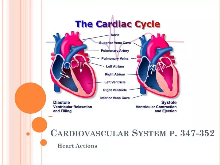
Cardiovascular System p. 347-352 Heart Actions
Cardiac Cycle • One complete heartbeat • Atrial contraction, ventricular relaxation • Atrial relaxation, ventricular contraction • Pressure within heart • Rises and falls as chambers fill and empty • Atrial or Ventricular Contraction • (Systole) Pressure • Pressure increases sharply • Atrial or Ventricular Relaxation • Diastole Pressure • Pressure goes back down • AV Valves are opening and closing during pressure changes
Heart Sounds • Vibrations in heart tissue produce opening and closing of valves • Heard as LubDup through stethoscope • Lub • Heard during ventricular contraction (AV valves are closing) • Dup • Heard during ventricular relaxation (pulmonary and aortic valves are shutting) • MurMur • Abnormal sound • Usually occurs when heart valves don’t properly close causing blood to leak through
Cardiac Muscle Fibers • Act like skeletal muscles but connect to a network of sending impulses to contract all as a unit. • Functional Syncytium • Mass of merging cells that act as a unit
Cardiac Conduction System • Coordinates events of cardiac cycle • Sinoatrial Node (SA Node) • Small mass of elongated specialized cardiac muscle tissue • Cells reach threshold on own and membranes contract one another • Initiate impulses that spread into the surrounding myocardium and stimulate cardiac muscle fiber to contract • Rhythmic activity • 70-80 impulses/min. (Pulse) • Pacemaker (Rhythmic contracting)
Atrioventricular Node (AV Node) • Located in septum • Receives slow impulse • After received impulse AV node sends it on to AV Bundle • Purkinje Fibers • Allow transmission of impulse for contraction of the ventricles and pushes blood on out to aorta
ECG/EKG…Electrocadiogram • Recording of the electrical changes in the myocardium during cardiac cycle • Deflections = waves on paper • Polarization and repolarization causes pen to move
P QRS T Waves • P wave • Depolarization, contraction of atriums • QRS Complex • Waves correspond to depolarization of ventriular fibers that contract ventricles, atriums relax, ventricles contract • T wave • Ventricles relax, pattern ends • P-R Interval • Wave travels through AV node, AV bundles, bundle branches and purkinje fibers • S-T Segment • Time for complete excitation of ventricles • Q-T Interval • Time required for complete excitation and recovery of ventricles • T-P Interlude • Time from completion of ventricular repolarization to next atrial excitation
Regulation of Cardiac Cycle • Read p. 356 • Arrhythmia • Abnormal heart rhythm • http://www.youtube.com/watch?v=xw4nDMgTOrw
Review • Describe the pressure changes in the atria and ventricles during a cardiac cycle. • What causes heart sounds? • What is a functional syncytium? • How is a cardiac impulse imitated? • How is a cardiac impulse transmitted from the right atrium to the other heart chambers?